In biology and ecology,abiotic components or abiotic factors are non-living chemical and physical parts of the environment that affect living organisms and the functioning of ecosystems. Abiotic factors and the phenomena associated with them underpin biology as a whole.
Abiotic components include physical conditions and non-living resources that affect living organisms in terms of growth,maintenance, and reproduction. Resources are distinguished as substances or objects in the environment required by one organism and consumed or otherwise made unavailable for use by other organisms.
Component degradation of a substance occurs by chemical or physical processes, e.g. hydrolysis. All non-living components of an ecosystem, such as atmospheric conditions and water resources, are called abiotic components. (W)
Abiotic factors are non living components found in an ecosystem which influence living things (biotic factors).
adaptive immune system
The adaptive immune system, also referred as the acquired immune system, is a subsystem of the immune system that is composed of specialized, systemic cells and processes that eliminates pathogens by preventing their growth. The acquired immune system is one of the two main immunity strategies found in vertebrates (the other being the innate immune system).
Acquired immunity creates immunological memory after an initial response to a specific pathogen, and leads to an enhanced response to subsequent encounters with that pathogen. This process of acquired immunity is the basis of vaccination. Like the innate system, the acquired system includes both humoral immunity components and cell-mediated immunity components. (W)
adenoviridae
Adenoviruses (members of the familyAdenoviridae) are medium-sized (90–100 nm),nonenveloped (without an outer lipid bilayer) viruses with an icosahedralnucleocapsid containing a double stranded DNA genome. Their name derives from their initial isolation from human adenoids in 1953.
They have a broad range of vertebrate hosts; in humans, more than 50 distinct adenoviral serotypes have been found to cause a wide range of illnesses, from mild respiratory infections in young children (known as the common cold) to life-threatening multi-organ disease in people with a weakened immune system.(W)
Transmission electron micrograph of two Adenovirus particles.
Schematic diagram of the linear adenovirus genome, showing Early genes (E) and Late genes (L).
The structure of adenovirus. 1 = penton capsomeres 2 = hexon capsomeres, and 3= viral genome (linear dsDNA).
It was used by the United States military from 1971 to 1999, but was discontinued when the only manufacturer stopped production. This vaccine elicited immunity to adenovirus serotypes 4 and 7, the serotypes most often associated with acute respiratory disease. On March 16, 2011, the U.S. Food and Drug Administration approved an adenovirus vaccine manufactured by Teva Pharmaceuticals under contract to the U.S. Army. This vaccine is essentially the same as the one used from 1971 to 1999. On October 24, 2011, the military services began administering the new adenovirus vaccine to recruits during basic training.
The vaccine is orally administered and consists of live (not attenuated) virus. The tablets are coated, so that the virus passes the stomach and infects the intestines, where the immune response is raised.
It should not be confused with the strategy of using adenovirus as a viral vector to develop vaccines for other pathogens, or as a general gene carrier.(W)
Extracted from the PDF. Shows two bottles of the TEVA adenovirus and monovirus vaccines made for the US Military.
adult stem cell
Adult stem cells are undifferentiatedcells, found throughout the body after development, that multiply by cell division to replenish dying cells and regenerate damaged tissues. Also known as somaticstem cells(from Greek Σωματικóς, meaning of the body), they can be found in juvenile as well as adult animals and humans, unlike embryonic stem cells.
Scientific interest in adult stem cells is centered on their ability to divide or self-renew indefinitely, and generate all the cell types of the organ from which they originate, potentially regenerating the entire organ from a few cells. Unlike for embryonic stem cells, the use of human adult stem cells in research and therapy is not considered to be controversial, as they are derived from adult tissue samples rather than human embryos designated for scientific research. They have mainly been studied in humans and model organisms such as mice and rats. (W)
Transmission electron micrograph of a mesenchymal stem cell displaying typical ultrastructural characteristics.
Stem cell division and differentiation. A – stem cells; B – progenitor cell; C – differentiated cell; 1 – symmetric stem cell division; 2 – asymmetric stem cell division; 3 – progenitor division; 4 – terminal differentiation.
Cyanidiophyceae is a class of unicellular red algae within division Cyanidiophytina, and contain a single plastid, one to three mitochondria, a nucleus, a vacuole and floridean starch. Most are extremophiles inhabiting acid hot springs.(W)
Gracilaria is a genus of red algae (Rhodophyta) notable for its economic importance as an agarophyte, as well as its use as a food for humans and various species of shellfish. (W)
allele (allel)
An allele (from Greek ἄλλος állos, "other") is a variant form of a given gene, meaning it is one of two or more versions of a known mutation at the same place on a chromosome. It can also refer to different sequence variations for a several-hundred base-pair or more region of the genome that codes for a protein. Alleles can come in different extremes of size. At the lowest possible end one can be the single base choice of a single nucleotide polymorphism (SNP). At the higher end, it can be the sequence variations for the regions of the genome that code for the same protein which can be up to several thousand base-pairs long. (W)
To elevate glucose levels, glucagon binds to receptors on hepatocytes (liver cells) and some other cells (e.g. kidney cells). This activates an enzyme, glycogen phosphorylase, inside the hepatocyte to hydrolyse glycogen to glucose. This process is called glycogenolysis. In rodents, alpha cells are located in the periphery of the islets, however in humans the islet architecture is generally less organized and alpha cells are frequently observed inside the islets as well. When being viewed by an electron microscope, alpha cells can be identified by their characteristic granules with a large dense core and a small white halo.
Alpha cells also generate GLP-1 and may have protective and regenerative effect on beta cells. They possibly can transdifferentiate into beta cells to replace lost beta cells. (W)
Islet of Langerhans, haemalum-eosin stain.
amoeba
An amoeba or ameba (plural am(o)ebas or am(o)ebae), often called an amoeboid, is a type of cell or unicellular organism which has the ability to alter its shape, primarily by extending and retracting pseudopods. Amoebae do not form a single taxonomic group; instead, they are found in every major lineage of eukaryotic organisms. Amoeboid cells occur not only among the protozoa, but also in fungi,algae, and animals.
Morphology of a naked amoeba in the genus Mayorella.
anchor cell
The anchor cell is a cell in nematodes such as Caenorhabditis elegans. It is important in the development of the reproductive system, as it is required for the production of the tube of cells that allows embryos to pass from the uterus through the vulva to the outside of the worm.
During the development of C. eleganshermaphrodites, the anchor cell produces a signalling molecule (LIN-3/EGF) that induces nearby epidermal cells to develop into the vulva. The anchor cell also produces another signal (the Notch ligand LAG-2) that induces adjacent uterine cells to become the π cells, some of which will later connect the uterus to the vulva. The anchor cell next removes the basement membrane that separates the uterus and vulva and invades, initiating the connection between the uterus and the vulva. Finally the anchor cell fuses with eight of the π cells to form the uterine seam cell.(W)
annular lipid shell
Annular lipids (also called shell lipids or boundary lipids) are a set of lipids or lipidic molecules which preferentially bind or stick to the surface of membrane proteins in biological cells. They constitute a layer, or an annulus/ shell, of lipids which are partially immobilized due to the existence of lipid-protein interactions. Polar headgroups of these lipids bind to the hydrophilic part of the membrane protein(s) at the inner and outer surfaces of lipid bilayer membrane. The hydrophobic surface of the membrane proteins is bound to the apposed lipid fatty acid chains of the membrane bilayer. For integral membrane proteins spanning the thickness of the membrane bilayer, these annular/shell lipids may act like a lubricating layer on the proteins' surfaces, thereby facilitating almost free rotation and lateral diffusion of membrane proteins within the 2-dimensional expanse of the biological membrane(s). Outside the layer of shell/annular lipids, lipids are not tied down to protein molecules. However, they may be slightly restricted in their segmental motion freedom due to mild peer pressure of protein molecules, if present in high concentration, which arises from extended influence of protein-lipid interaction. Membrane areas away from protein molecules contain lamellar phase bulk lipids, which are largely free from any restraining effects due to protein-lipid interactions. Thermal denaturation of membrane proteins may destroy the secondary and tertiary structure of membrane proteins, exposing newer surfaces to membrane lipids and therefore increasing the number of lipids molecules in the annulus/shell layer. This phenomenon can be studied by the spin labelelectron paramagnetic resonance technique. (W)
Almost all cell types can present antigens in some way. They are found in a variety of tissue types. Professional antigen-presenting cells, including macrophages,B cells and dendritic cells, present foreign antigens to helper T cells, while virus-infected cells (or cancer cells) can present antigens originating inside the cell to cytotoxic T cells. In addition to the MHC family of proteins, antigen presentation relies on other specialized signaling molecules on the surfaces of both APCs and T cells.
Antigen-presenting cells are vital for effective adaptive immune response, as the functioning of both cytotoxic and helper T cells is dependent on APCs. Antigen presentation allows for specificity of adaptive immunity and can contribute to immune responses against both intracellular and extracellular pathogens. It is also involved in defense against tumors. Some cancer therapies involve the creation of artificial APCs to prime the adaptive immune system to target malignant cells. (W)
Antigen presentation stimulates immature T cells to become either mature "cytotoxic" CD8+ cells or mature "helper" CD4+ cells.
Antigen presentation stimulates T cells to activate "cytotoxic" CD8+ cells or "helper" CD4+ cells. Cytotoxic cells directly attack other cells carrying certain foreign or abnormal molecules on their surfaces. Helper T cells, or Th cells, coordinate immune responses by communicating with other cells. In most cases, T cells only recognize an antigen if it is carried on the surface of a cell by one of the body’s own MHC, or major histocompatibility complex, molecules.
The sub images where extracted from a time-lapse microscopy video showing apoptosis of DU145 prostate cancer cells. To induce apoptosis the cells were treated with etoposide. The 61 hour time-lapse was created using the HoloMonitor M3 from Phase Holographic Imaging AB.
Overview of signal transduction pathways.
Overview of TNF signalling in apoptosis, an example of direct signal transduction..
Overview of Fas signalling in apoptosis, an example of direct signal transduction.
Archaeal cells have unique properties separating them from the other two domains of life,Bacteria and Eukaryota. Archaea are further divided into multiple recognized phyla. Classification is difficult because most have not been isolated in a laboratory and have been detected only by their gene sequences in environmental samples.
Archaea are a major part of Earth's life. They are part of the microbiota of all organisms. In the human microbiota, they are important in the gut, mouth, and on the skin. Their morphological, metabolic, and geographical diversity permits them to play multiple ecological roles: carbon fixation; nitrogen cycling; organic compound turnover; and maintaining microbial symbiotic and syntrophic communities, for example. (W)
Modified drawing of the neural circuitry of the rodent hippocampus.
The archicortex in humans is a synonym of the hippocampal formation. The hippocampal formation is shown here, as drawn by Santiago Ramon y Cajal: DG: dentate gyrus. Sub: subiculum. EC: entorhinal cortex. CA1-CA3: hippocampus proper.
From: Santiago Ramón y Cajal (1911) [1909] Histologie du Système nerveux de l'Homme et des Vertébrés, Paris: A. Maloine.
An asymmetric cell division produces two daughter cells with different cellular fates. This is in contrast to symmetric cell divisions which give rise to daughter cells of equivalent fates. Notably, stem cells divide asymmetrically to give rise to two distinct daughter cells: one copy of the original stem cell as well as a second daughter programmed to differentiate into a non-stem cell fate. (In times of growth or regeneration, stem cells can also divide symmetrically, to produce two identical copies of the original cell.)
In principle, there are two mechanisms by which distinct properties may be conferred on the daughters of a dividing cell. In one, the daughter cells are initially equivalent but a difference is induced by signaling between the cells, from surrounding cells, or from the precursor cell. This mechanism is known as extrinsic asymmetric cell division. In the second mechanism, the prospective daughter cells are inherently different at the time of division of the mother cell. Because this latter mechanism does not depend on interactions of cells with each other or with their environment, it must rely on intrinsic asymmetry. The term asymmetric cell division usually refers to such intrinsic asymmetric divisions. (W)
Asymmetric cell division is integral during development. In spiralia, the first cleavage can be either symmetric or asymmetric, as shown in the left panel. Asymmetry can be accomplished through simple unequal segregation of cell fate determinants across a single plane, through sequestration of cell fate determinants in a polar lobe which is absorbed by one of the daughter cells, or a combination of both processes. The right panel summarizes the mechanisms of spiralian asymmetric cleavage discussed here. Red features indicate the molecule(s) implicated in establishing asymmetry.
autophagy
Autophagy (or autophagocytosis) (from the Ancient Greekαὐτόφαγοςautóphagos, meaning "self-devouring" and κύτοςkýtos, meaning "hollow") is the natural, regulated mechanism of the cell that removes unnecessary or dysfunctional components. It allows the orderly degradation and recycling of cellular components.(W)
(A) Diagram of the process of autophagy, which produces the structures autophagosomes, AP, and autolysomes, AL; (B) Electron micrograph of autophagic structures AP and AL in the fatbody of a fruit fly larva; (C) Fluorescently labeled autophagosomes AP in liver cells of starved mice.
autotroph (kendibeslek) (heterotroph: yanbeslek)
An autotroph or primary producer is an organism that produces complex organic compounds (such as carbohydrates,fats, and proteins) using carbon from simple substances such as carbon dioxide, generally using energy from light (photosynthesis) or inorganic chemical reactions (chemosynthesis). Autotrophs do not need a living source of carbon or energy and are the producers in a food chain, such as plants on land or algae in water (in contrast to heterotrophs as consumers of autotrophs or other heterotrophs). Autotrophs can reduce carbon dioxide to make organic compounds for biosynthesis and as stored chemical fuel. Most autotrophs use water as the reducing agent, but some can use other hydrogen compounds such as hydrogen sulfide. (W)
Flowchart to determine if a species is autotroph, heterotroph, or a subtype.
axon
An axon (from Greek ἄξων áxōn, axis), or nerve fiber (or nervefibre: see spelling differences) , is a long, slender projection of a nerve cell, or neuron, in vertebrates, that typically conducts electrical impulses known as action potentials away from the nerve cell body. The function of the axon is to transmit information to different neurons, muscles, and glands. In certain sensory neurons (pseudounipolar neurons) , such as those for touch and warmth, the axons are called afferent nerve fibers and the electrical impulse travels along these from the periphery to the cell body, and from the cell body to the spinal cord along another branch of the same axon. Axon dysfunction has caused many inherited and acquired neurological disorders which can affect both the peripheral and central neurons. Nerve fibers are classed into three types – group A nerve fibers,group B nerve fibers, and group C nerve fibers. Groups A and B are myelinated, and group C are unmyelinated. These groups include both sensory fibers and motor fibers. Another classification groups only the sensory fibers as Type I, Type II, Type III, and Type IV. (W)
Multipolar Neuron.
Detail showing microtubules at axon hillock and initial segment.
Transmission electron micrograph of a human B cell.
bacterial conjugation
Bacterial conjugation is the transfer of genetic material between bacterial cells by direct cell-to-cell contact or by a bridge-like connection between two cells. This takes place through a pilus. It is a parasexual mode of reproduction in bacteria.
Classical E. coli bacterial conjugation is often regarded as the bacterial equivalent of sexual reproduction or mating since it involves the exchange of genetic material. However, it is not sexual reproduction, since no exchange of gamete occurs, and indeed no generation of a new organism: instead an existing organism is transformed. During classical E. coli conjugation the donor cell provides a conjugative or mobilizable genetic element that is most often a plasmid or transposon. Most conjugative plasmids have systems ensuring that the recipient cell does not already contain a similar element.
The genetic information transferred is often beneficial to the recipient. Benefits may include antibiotic resistance,xenobiotic tolerance or the ability to use new metabolites. Others elements can be detrimental and may be viewed as bacterial parasites.
Conjugation in Escherichia coli by spontaneous zygogenesis[6] and in Mycobacterium smegmatis by distributive conjugal transfer differ from the more well studied classical E. coli conjugation in that these cases involve substantial blending of the parental genomes. (W)
Schematic drawing of bacterial conjugation.
Schematic drawing of bacterial conjugation. Conjugation diagram 1- Donor cell produces pilus. 2- Pilus attaches to recipient cell, brings the two cells together. 3- The mobile plasmid is nicked and a single strand of DNA is then transferred to the recipient cell. 4- Both cells recircularize their plasmids, synthesize second strands, and reproduce pili; both cells are now viable donors.
1.The insertion sequences (yellow) on both the F factor plasmid and the chromosome have similar sequences, allowing the F factor to insert itself into the genome of the cell. This is called homologous recombination and creates an Hfr (high frequency of recombination) cell. 2.The Hfr cell forms a pilus and attaches to a recipient F- cell. 3.A nick in one strand of the Hfr cell's chromosome is created. 4.DNA begins to be transferred from the Hfr cell to the recipient cell while the second strand of its chromosome is being replicated. 5.The pilus detaches from the recipient cell and retracts. The Hfr cell ideally wants to transfer its entire genome to the recipient cell. However, due to its large size and inability to keep in contact with the recipient cell, it is not able to do so. 6.a. The F- cell remains F- because the entire F factor sequence was not received. Since no homologous recombination occurred, the DNA that was transferred is degraded by enzymes. b. In very rare cases, the F factor will be completely transferred and the F- cell will become an Hfr cell.
bacteriophage
A bacteriophage, also known informally as a phage, is a virus that infects and replicates within bacteria and archaea. The term was derived from "bacteria" and the Greek φαγεῖν (phagein), meaning "to devour". Bacteriophages are composed of proteins that encapsulate a DNA or RNAgenome, and may have structures that are either simple or elaborate. Their genomes may encode as few as four genes (e.g. MS2) and as many as hundreds of genes. Phages replicate within the bacterium following the injection of their genome into its cytoplasm.
Bacteriophages are among the most common and diverse entities in the biosphere. Bacteriophages are ubiquitous viruses, found wherever bacteria exist. It is estimated there are more than 1031 bacteriophages on the planet, more than every other organism on Earth, including bacteria, combined. One of the densest natural sources for phages and other viruses is seawater, where up to 9x10⁸ virions per millilitre have been found in microbial mats at the surface, and up to 70% of marine bacteria may be infected by phages.
Phages have been used since the late 20th century as an alternative to antibiotics in the former Soviet Union and Central Europe, as well as in France. They are seen as a possible therapy against multi-drug-resistant strains of many bacteria (see phage therapy). On the other hand, phages of Inoviridae have been shown to complicate biofilms involved in pneumonia and cystic fibrosis and to shelter the bacteria from drugs meant to eradicate disease, thus promoting persistent infection. (W)
Atomic structural model of bacteriophage t4. By Victor Padilla-Sanchez, PhD (Washington DC).
The structure of a typical myovirus bacteriophage.
Artistic rendering of a T4 bacteriophage. The colours grey and orange do not signify anything, they are just used to illustrate structure. Created for Wikipedia.
Anatomy and infection cycle of phage T4.
Anatomy and infection cycle of phage T4. 1: Attachment of phage's fibres to a bacterium, 2: Injection of DNA, 3: Synthesis of phage components, 4: Assembly of new phages, 5: Burst of bacterium and release of infectious phages..
Bacteriophage P22, a member of the Podoviridae by morphology due to its short, non-contractile tail.
P22 is genetically a member of the lambda-like bacteriophage family, and morphologically a member of the Podoviridae. This electron micrograph is by Sherwood Casjens and Elaine Lenk. Sherwood Casjens granted permission for the use of the image at wikipedia.org. Crenim (talk) 16:36, 8 September 2011 (UTC).
Diagram of the DNA injection process.
The injection process of bacteriophage DNA into a bacterial cell .
In this electron micrograph of bacteriophages attached to a bacterial cell, the viruses are the size and shape of coliphage T1.
Transmission electron micrograph of multiple bacteriophages attached to a bacterial cell wall; the magnification is approximately 200,000.
bacterivore
Bacterivores are free-living, generally heterotrophic organisms, exclusively microscopic, which obtain energy and nutrients primarily or entirely from the consumption of bacteria. Many species of amoeba are bacterivores, as well as other types of protozoans. Commonly, all species of bacteria will be prey, but spores of some species, such as Clostridium perfringens, will never be prey, because of their cellular attributes. (W)
basement membrane (Lat. membrana basalis)
The basement membrane is a thin, pliable sheet-like type of extracellular matrix, that provides cell and tissue support and acts as a platform for complex signalling. The basement membrane sits between epithelial tissues including mesothelium and endothelium, and the underlying connective tissue.
To represent the above in a visually organised manner, the basement membrane is organized as follows:
Beta cells (β cells) are a type of cell found in pancreatic islets that synthesize and secrete insulin and amylin. Beta cells make up 50–70% of the cells in human islets. In patients with Type I Diabetes, beta-cell mass and function are diminished, leading to insufficient insulin secretion and hyperglycemia. (W)
The Consensus Model of glucose-stimulated insulin secretion.
Cross-sectional view of the structures that can be formed by phospholipids in an aqueous solution.
Cross section of the different structures that phospholipids can take in a aqueous solution. The circles are the hydrophilic heads and the wavy lines are the fatty acyl side chains.
A fluid membrane model of the phospholipid bilayer.
Caption: The cell membrane of the cell is a phospholipid bilayer containing many different molecular components, including proteins and cholesterol, some with carbohydrate groups attached. URL: https://cnx.org/contents/FPtK1zmh@8.108:q2X995E3@12/The-Cell-Membrane Version 8.25 from the Textbook OpenStax Anatomy and Physiology Published May 18, 2016
biome
A biome is a community of plants and animals that have common characteristics for the environment they exist in. They can be found over a range of continents. Biomes are distinct biological communities that have formed in response to a shared physical climate.Biome is a broader term than habitat; any biome can comprise a variety of habitats. (W)
One way of mapping terrestrial biomes around the world.
blood-brain barrier
The blood–brain barrier (BBB) is a highly selective semipermeable border of endothelial cells that prevents solutes in the circulating blood from non-selectively crossing into the extracellular fluid of the central nervous system where neurons reside. The blood-brain barrier is formed by endothelial cells of the capillary wall,astrocyte end-feet ensheathing the capillary, and pericytes embedded in the capillary basement membrane. This system allows the passage of some molecules by passive diffusion, as well as the selective and active transport of various nutrients, ions, organic anions, and macromolecules such as glucose, water and amino acids that are crucial to neural function.
The blood-brain barrier restricts the passage of pathogens, the diffusion of solutes in the blood, and large or hydrophilic molecules into the cerebrospinal fluid, while allowing the diffusion of hydrophobic molecules (O2, CO2, hormones) and small polar molecules. Cells of the barrier actively transport metabolic products such as glucose across the barrier using specific transport proteins. The barrier also restricts the passage of peripheral immune factors, like signaling molecules, antibodies, and immune cells, into the CNS, thus insulating the brain from damage due to peripheral immune events.
Blood-brain barrier.
Protective barriers of the brain. The collective term “blood-brain barrier” is used to describe four main interfaces between the central nervous system and the periphery. (i) The blood-brain barrier proper formed by tight junctions between the endothelial cells of the cerebral vasculature. It is thought that pericytes (peri.) are sufficient to induce some barrier characteristics in endothelial cells, while astrocytes (astro.) are able to maintain the integrity of the blood-brain barrier postnatally. (ii) The blood-CSF barrier formed by tight junctions between epithelial cells of the choroid plexus epithelial cells (note the plexus vasculature is fenestrated). Resident epiplexus (epi.) immune cells are present on the CSF-surface of the plexus epithelium. (iii) The outer CSF-brain barrier and the level of the pia arachnoid, formed by tight junctions between endothelial cells of the arachnoid vessels. (iv) The inner CSF-brain barrier, present only in early development, formed by strap junctions between the neuroependymal cells lining the ventricular surfaces. In the adult this barrier is no longer present. Both the blood-brain and CSF-brain barriers extend down the spinal cord. The CSF-filled ventricular system is depicted in blue, while CNS brain tissue is in brown. The lateral ventricular choroid plexuses are shown in red. Abbreviations: astro, astrocyte; bv, blood vessel; cpec, choroid plexus epithelial cell; csf, cerebrospinal fluid; peri, pericytes. (W)
Part of a network of capillaries supplying brain cells.
A network of capillaries supply brain cells with nutrients. Tight seals in their walls keep blood toxins—and many beneficial drugs—out of the brain. From: Bridging the Blood-Brain Barrier: New Methods Improve the Odds of Getting Drugs to the Brain Cells That Need Them Ferber D PLoS Biology Vol. 5, No. 6, e169 doi:10.1371/journal.pbio.0050169.
The astrocytes type 1 surrounding capillaries in the brain.
The Blood Brain Barrier and Astrocytes type 1 .
Sketch showing constitution of blood vessels inside the brain.
blood plasma
Blood plasma is a yellowish liquid component of blood that holds the blood cells of whole blood in suspension. It is the liquid part of the blood that carries cells and proteins throughout the body. It makes up about 55% of the body's total blood volume. It is the intravascular fluid part of extracellular fluid (all body fluid outside cells). It is mostly water (up to 95% by volume), and contains important dissolved proteins (6–8%) (e.g., serum albumins,globulins, and fibrinogen),glucose,clotting factors,electrolytes (Na+, Ca2+, Mg2+, HCO3-, Cl-, etc.), hormones,carbon dioxide (plasma being the main medium for excretory product transportation), and oxygen. It plays a vital role in an intravascular osmotic effect that keeps electrolyte concentration balanced and protects the body from infection and other blood disorders. (W)
Body fluids, bodily fluids, or biofluids are liquids within the human body. In lean healthy adult men, the total body water is about 60% (60–67%) of the total body weight; it is usually slightly lower in women. The exact percentage of fluid relative to body weight is inversely proportional to the percentage of body fat. A lean 70 kg (160 pound) man, for example, has about 42 (42–47) liters of water in his body.
The total body of water is divided between the intracellular fluid (ICF) compartment (also called space, or volume) and the extracellular fluid (ECF) compartment (space, volume) in a two-to-one ratio: 28 (28–32) liters are inside cells and 14 (14–15) liters are outside cells.
The ECF compartment is divided into the interstitial fluid volume – the fluid outside both the cells and the blood vessels – and the intravascular volume (also called the vascular volume and blood plasma volume) – the fluid inside the blood vessels – in a three-to-one ratio: the interstitial fluid volume is about 12 liters, the vascular volume is about 4 liters.
The interstitial fluid compartment is divided into the lymphatic fluid compartment – about 2/3's, or 8 (6–10) liters; the transcellular fluid compartment is the remaining 1/3, or about 4 liters.
The vascular volume is divided into the venous volume and the arterial volume; and the arterial volume has a conceptually useful but unmeasurable subcompartment called the effective arterial blood volume.(W)
Intracellular and extracellular fluid compartments. The extracellular fluid compartment is further subdivided into the interstitial fluid and the intravascular fluid compartments.
body water
In physiology,body water is the water content of an animal body that is contained in the tissues, the blood, the bones and elsewhere. The percentages of body water contained in various fluid compartments add up to total body water (TBW). This water makes up a significant fraction of the human body, both by weight and by volume. Ensuring the right amount of body water is part of fluid balance, an aspect of homeostasis.(W)
c
cap formation
When molecules on the surface of cell are crosslinked, they are moved to one end of the cell to form a "cap". This phenomenon, the process of which is called cap formation, was discovered in 1971 on lymphocytes and is a property of amoebae and all locomotory animal cells except sperm. The crosslinking is most easily achieved using a polyvalent antibody to a surface antigen on the cell. Cap formation can be visualised by attaching a fluorophore, such as fluorescein, to the antibody. (W)
carcinogenesis
Carcinogenesis, also called oncogenesis or tumorigenesis, is the formation of a cancer, whereby normal cells are transformed into cancer cells. The process is characterized by changes at the cellular, genetic, and epigenetic levels and abnormal cell division. Cell division is a physiological process that occurs in almost all tissues and under a variety of circumstances. Normally the balance between proliferation and programmed cell death, in the form of apoptosis, is maintained to ensure the integrity of tissues and organs. According to the prevailing accepted theory of carcinogenesis, the somatic mutation theory, mutations in DNA and epimutations that lead to cancer disrupt these orderly processes by disrupting the programming regulating the processes, upsetting the normal balance between proliferation and cell death. This results in uncontrolled cell division and the evolution of those cells by natural selection in the body. Only certain mutations lead to cancer whereas the majority of mutations do not. (W)
Cancers and tumors are caused by a series of mutations. Each mutation alters the behavior of the cell somewhat.
cell
The cell (Latin cella, meaning "small room") is the basic structural, functional, and biological unit of all known organisms. A cell is the smallest unit of life. Cells are often called the "building blocks of life". The study of cells is called cell biology, cellular biology, or cytology. (W)
The cell (from Latincella, meaning "small room") is the basic structural, functional, and biological unit of all known organisms. A cell is the smallest unit of life. Cells are often called the "building blocks of life". The study of cells is called cell biology, cellular biology, or cytology.
The number of cells in plants and animals varies from species to species; it has been estimated that humans contain somewhere around 40 trillion (4×10¹³) cells. The human brain accounts for around 80 billion of these cells.
Cells were discovered by Robert Hooke in 1665, who named them for their resemblance to cells inhabited by Christian monks in a monastery. Cell theory, first developed in 1839 by Matthias Jakob Schleiden and Theodor Schwann, states that all organisms are composed of one or more cells, that cells are the fundamental unit of structure and function in all living organisms, and that all cells come from pre-existing cells. Cells emerged on Earth at least 3.5 billion years ago.
The plasmodesmata, pores in the cell wall that link adjacent cells and allow plant cells to communicate with adjacent cells. Animals have a different but functionally analogous system of gap junctions between adjacent cells.
Bryophytes and seedless vascular plants only have flagellae and centrioles in the sperm cells. Sperm of cycads and Ginkgo are large, complex cells that swim with hundreds to thousands of flagellae.
📹 Campbell’s Biology — A Tour of the Cell / Peer Vids (LINK)
cell adhesionCell adhesion is the process by which cells interact and attach to neighbouring cells through specialised molecules of the cell surface. This process can occur either through direct contact between cell surfaces such as cell junctions or indirect interaction, where cells attach to surrounding extracellular matrix, a gel-like structure containing molecules released by cells into spaces between them. Cells adhesion occurs from the interactions between cell-adhesion molecules (CAMs), transmembrane proteins located on the cell surface. Cell adhesion links cells in different ways and can be involved in signal transduction for cells to detect and respond to changes in the surroundings. Other cellular processes regulated by cell adhesion include cell migration and tissue development in multicellular organisms. Alterations in cell adhesion can disrupt important cellular processes and lead to a variety of diseases, including cancer and arthritis. Cell adhesion is also essential for infectious organisms, such as bacteria or viruses, to cause diseases. (W)
Schematic of cell adhesion.
Overview diagram of different types of cell junctions present in epithelial cells, including cell–cell junctions and cell–matrix junctions.
Adheren junction showing homophilic binding between cadherins and how catenin links it to actin filaments.
Gap junctions showing connexons and connexins.
Hemidesmosomes diagram showing interaction between integrins and laminin, including how integrins are linked to keratin intermediate filaments.
The cell cycle, or cell-division cycle, is the series of events that take place in a cell that cause it to divide into two daughter cells. These events include the duplication of its DNA (DNA replication) and some of its organelles, and subsequently the partitioning of its cytoplasm and other components into two daughter cells in a process called cell division.(W)
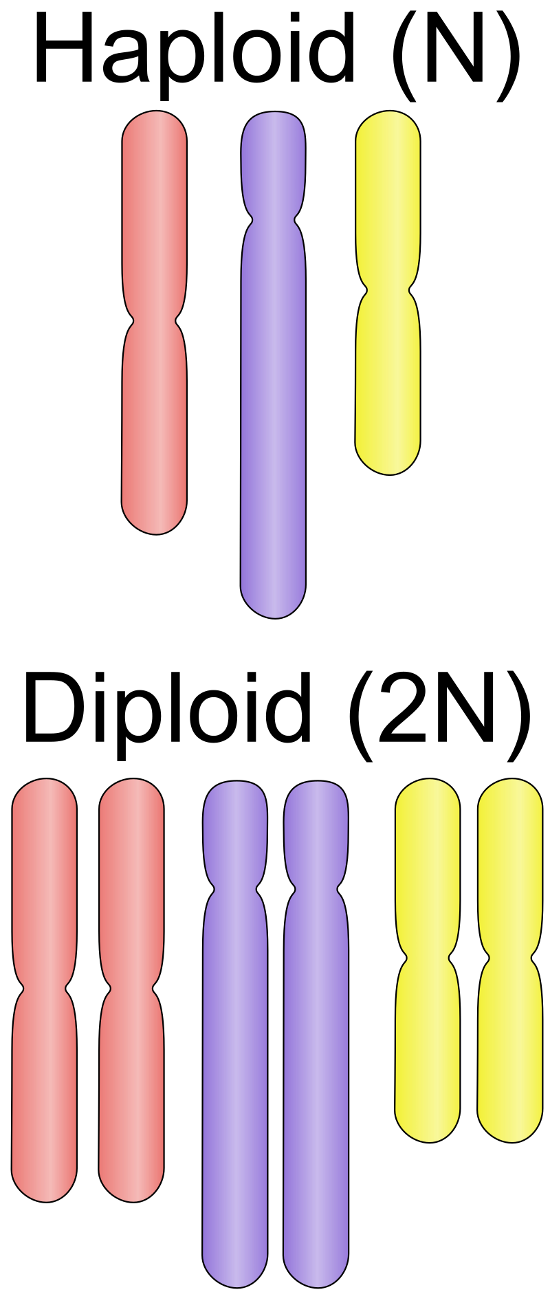
Cell division is the process by which a parent cell divides into two or more daughter cells. Cell division usually occurs as part of a larger cell cycle. In eukaryotes, there are two distinct types of cell division; a vegetative division, whereby each daughter cell is genetically identical to the parent cell (mitosis), and a reproductive cell division, whereby the number of chromosomes in the daughter cells is reduced by half to produce haploid gametes (meiosis). In cell biology,mitosis is a part of the cell cycle, in which, replicated chromosomes are separated into two new nuclei. Cell division gives rise to genetically identical cells in which the total number of chromosomes is maintained. In general, mitosis (division of the nucleus) is preceded by the S stage of interphase (during which the DNA is replicated) and is often followed by telophase and cytokinesis; which divides the cytoplasm,organelles and cell membrane of one cell into two new cells containing roughly equal shares of these cellular components. The different stages of Mitosis all together define the mitotic (M) phase of an animal cell cycle—the division of the mother cell into two daughter cells genetically identical to each other. Meiosis results in four haploid daughter cells by undergoing one round of DNA replication followed by two divisions. Homologous chromosomes are separated in the first division, and sister chromatids are separated in the second division. Both of these cell division cycles are used in the process of sexual reproduction at some point in their life cycle. Both are believed to be present in the last eukaryotic common ancestor. (W)
Cell division in prokaryotes (binary fission) and eukaryotes (mitosis and meiosis).
Vectorised version of File:Three cell growth types.png. Three types of cell reproduction are compared: the relatively simple Binary fission and two more complicated types that either involve mitosis or meiosis.Binary fission. Organisms such as bacteria typically have a single chromosome (green). At the start of the binary fission process, the DNA molecule of the cell's chromosome is replicated, producing two copies of the chromosome. A key aspect of bacterial cell reproduction is making sure that each daughter cell gets a copy of the chromosome. Cytokinesis is the actual physical separation of the two new daughter cells. Cell reproduction that involves mitosis. Most eukaryotic organisms like humans have more than one chromosome. In order to make sure that a copy of each chromosome gets segregated into each daughter cell, the spindle apparatus is used (blue threads). The chromosomes are moved along the long thin microtubules like trains moving along train tracks. Humans are diploid; we have two copies of each type of chromosome, one from the father (red) and one from the mother (green). Cell reproduction that involves meiosis. The human sex cells (gametes) are produced by meiosis. For sperm production there are two cytokinesis steps that produce a total of four cells, each with half the normal number of chromosomes. The situation is different in the ovaries for egg production where one of the four sets of chromosomes that is segregated is placed in a large egg cell, ready to be combined with the DNA from a sperm cell (see meiosis for details). Note text is on a hidden layer under the visible text for consistent fonts. (W).
Divisome and elongasome complexes responsible for peptidoglycan synthesis during lateral cell-wall growth and division.
a Cells of the same lineage fuse to form a cell with multiple nuclei, known as a syncytium. The fused cell can have an altered phenotype and new functions such as barrier formation. b Cells of different lineage fuse to form a cell with multiple nuclei, known as a heterokaryon. The fused cells might have undergone a reversion of phenotype or show transdifferentiation. c Cells of different lineage or the same lineage fuse to form a cell with a single nucleus, known as a synkaryon. New functions of the fused cell can include a reversion of phenotype, transdifferentiation and proliferation. If nuclear fusion occurs, the fused nucleus initially contains the complete chromosomal content of both fusion partners (4N), but ultimately chromosomes are lost and/or re-sorted (see arrows). If nuclear fusion does not occur, a heterokaryon (or syncytium) can become a synkaryon by shedding an entire nucleus.
Cell junctions are also especially important in enabling communication between neighboring cells via specialized protein complexes called communicating (gap) junctions. Cell junctions are also important in reducing stress placed upon cells.
Cell membranes are a mosaic of protein and lipid molecules, both of which can drift from place to place within the membrane. Most of the surface area of the membrane consists of a type of lipid called phospholipid. Each phospholipid molecule has a polar head and a nonpolar tail. The phosphate-containing head is hydrophilic, or “water-loving” because its polar structure is attached to water molecules, which are also polar. The fatty acid tail is hydrophobic, because it is nonpolar and repels water molecules. Cell membranes are phospholipid bilayers, a double-layered structure in which the hydrophilic heads of the phospholipids face outward. The middle of the membrane contains the hydrophobic tails of the phospholipids, an arrangement that shields them from contact with water molecules. Whereas membrane lipids act as a general barrier around the cell, membrane proteins have specific functions. For example, they may act as enzymes, receptors, anchors, or channels. There are two major populations of membrane proteins: integral proteins, which are part of the membrane structure and usually span the width of both phospholipid layers, and peripheral proteins, which are bound to the surfaces of the membrane.
Due to the highly viscous environment (low Reynolds number), cells need to continuously produce forces in order to move. Cells achieve active movement by very different mechanisms. Many less complex prokaryotic organisms (and sperm cells) use flagella or cilia to propel themselves. Eukaryotic cell migration typically is far more complex and can consist of combinations of different migration mechanisms. It generally involves drastic changes in cell shape which are driven by the cytoskeleton. Two very distinct migration scenarios are crawling motion (most commonly studied) and blebbing motility. A paradigmatic example of crawling motion is the case of fish epidermal keratocytes, which have been extensively used in research and teaching. (W)
Two different models for how cells move. A) Cytoskeletal model. B) Membrane Flow Model.
(A) Dynamic microtubules are necessary for tail retraction and are distributed at the rear end in a migrating cell. Green, highly dynamic microtubules; yellow, moderately dynamic microtubules and red, stable microtubules. (B) Stable microtubules act as struts and prevent tail retraction and thereby inhibit cell migration..
Rearward membrane flow (red arrows) and vesicle trafficking from back to front (blue arrows) drive adhesion-independent migration.
Schematic representation of the collective biomechanical and molecular mechanism of cell motion.
cell nucleus
HeLa cells stained for nuclear DNA with the blue fluorescentHoechst dye. The central and rightmost cell are in interphase, thus their entire nuclei are labeled. On the left, a cell is going through mitosis and its DNA has condensed.
A mouse fibroblast nucleus in which DNA is stained blue. The distinct chromosome territories of chromosome 2 (red) and chromosome 9 (green) are stained with fluorescent in situ hybridization.
3D rendering of nucleus with location of nucleolus.
The eukaryotic cell nucleus. Visible in this diagram are the ribosome-studded double membranes of the nuclear envelope, the DNA (complexed as chromatin) , and the nucleolus. Within the cell nucleus is a viscous liquid called nucleoplasm, similar to the cytoplasm found outside the nucleus..
cell polarity
Cell polarity refers to spatial differences in shape, structure, and function within a cell. Almost all cell types exhibit some form of polarity, which enables them to carry out specialized functions. Classical examples of polarized cells are described below, including epithelial cells with apical-basal polarity, neurons in which signals propagate in one direction from dendrites to axons, and migrating cells. Furthermore, cell polarity is important during many types of asymmetric cell division to set up functional asymmetries between daughter cells.
Many of the key molecular players implicated in cell polarity are well conserved. For example, in metazoan cells, the PAR-3/PAR-6/aPKC complex plays a fundamental role in cell polarity. While the biochemical details may vary, some of the core principles such as negative and/or positive feedback between different molecules are common and essential to many known polarity systems. (W)
Polarized localization of Staufen protein (white arrow) in Drosophila stage 9 oocyte (Stau:GFP, DAPI).
cell potency
Cell potency is a cell's ability to differentiate into other cell types. The more cell types a cell can differentiate into, the greater its potency. Potency is also described as the gene activation potential within a cell, which like a continuum, begins with totipotency to designate a cell with the most differentiation potential, pluripotency,multipotency,oligopotency, and finally unipotency.(W)
A: Human embryonic stem cells (cell colonies that are not yet differentiated). B: Nerve cells.
(A) Human Embryonic Stem Cells (hESCs); (B) neurons derived from hESCs.
Naive human pluripotent stem cell colony here seen growing on feeder cells (mouse).
Hematopoietic stem cells are an example of multipotency. When they differentiate into myeloid or lymphoid progenitor cells, they lose potency and become oligopotent cells with the ability to give rise to all cells of its lineage.
This diagram shows the hematopoiesis as it occurs in humans. It may look incomplete when rendered directly from WikiMedia. Reference list is found at: File:Hematopoiesis (human) diagram.png The morphological characteristics of the hematopoietic cells are shown as seen in a Wright’s stain, May-Giemsa stain or May-Grünwald-Giemsa stain. Alternative names of certain cells are indicated between parentheses. Certain cells may have more than one characteristic appearance. In these cases, more than one representation of the same cell has been included. Together, the monocyte and the lymphocytes comprise the agranulocytes, as opposed to the granulocytes (basophil, neurtophil and eosinophil) that are produced during granulopoiesis. B., N. and E. stand for Basophilic, Neutrophilic and Eosinophilic, respectively – as in Basophilic promyelocyte. For lymphocytes, the T and B are actual designations. [1] The polychromatic erythrocyte (reticulocyte) at the right shows its characteristic appearance when stained with methylene blue or Azure B. [2] The erythrocyte at the right is a more accurate representation of its appearance in reality when viewed through a microscope. [3] Other cells that arise from the monocyte: osteoclast, microglia (central nervous system), Langerhans cell (epidermis), Kupffer cell (liver). [4] For clarity, the T and B lymphocyte are split to better indicate that the plasma cell arises from the B-cell. Note that there is no difference in the appearance of B- and T-cells unless specific staining is applied.
cell signaling
n biology,cell signaling (cell signalling in British English) is part of any communication process that governs basic activities of cells and coordinates multiple-cell actions. A signal is an entity that codes or conveys information. Biological processes are complex molecular interactions that involve a lot of signals. The ability of cells to perceive and correctly respond to their microenvironment is the basis of development,tissue repair, and immunity, as well as normal tissue homeostasis. Errors in signaling interactions and cellular information processing may cause diseases such as cancer,autoimmunity, and diabetes. By understanding cell signaling, clinicians may treat diseases more effectively and, theoretically, researchers may develop artificial tissues.(W)
External reactions and internal reactions for signal transduction (click to enlarge).
Three conformation states of acetylcholine receptor (click to enlarge).
Sketch of an enzyme-linked receptor structure (structure of IGF-1R) (click to enlarge).
Flow charts of two strategies of structure-based drug design.
cell wall
A cell wall is a structural layer surrounding some types of cells, just outside the cell membrane. It can be tough, flexible, and sometimes rigid. It provides the cell with both structural support and protection, and also acts as a filtering mechanism. Cell walls are present in most prokaryotes (except mollicute bacteria), in algae,fungi and eukaryotes including plants but are absent in animals. A major function is to act as pressure vessels, preventing over-expansion of the cell when water enters.
Cellular differentiation is the process in which a cell changes from one cell type to another. Usually, the cell changes to a more specialized type. Differentiation occurs numerous times during the development of a multicellular organism as it changes from a simple zygote to a complex system of tissues and cell types. Differentiation continues in adulthood as adult stem cells divide and create fully differentiated daughter cells during tissue repair and during normal cell turnover. Some differentiation occurs in response to antigen exposure. Differentiation dramatically changes a cell's size, shape, membrane potential,metabolic activity, and responsiveness to signals. These changes are largely due to highly controlled modifications in gene expression and are the study of epigenetics. With a few exceptions, cellular differentiation almost never involves a change in the DNA sequence itself. Thus, different cells can have very different physical characteristics despite having the same genome.(W)
Mechanisms of cellular differentiation.
Stem cell differentiation into various tissue types.
An overview of major signal transduction pathways.
Nuclear Reprogramming methods: SCNT, Direct Reprogramming.
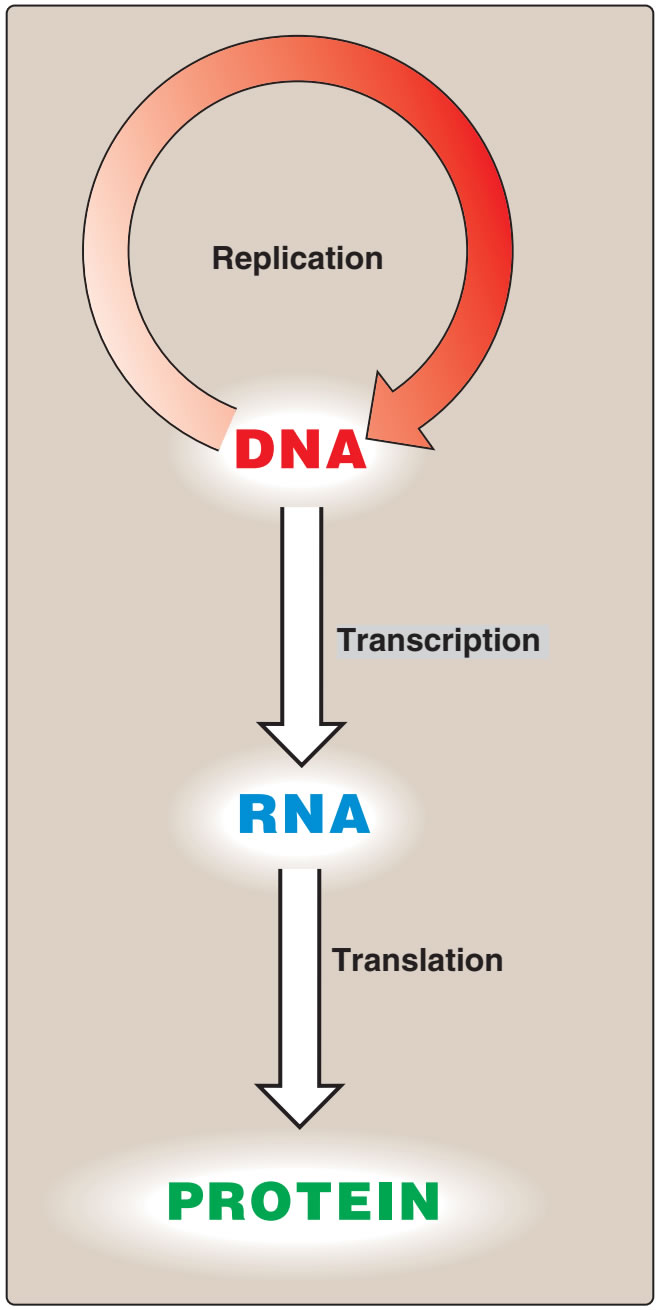
central dogma of molecular biology
The central dogma of molecular biology is an explanation of the flow of genetic information within a biological system. It is often stated as "DNA makes RNA, and RNA makes protein", although this is not its original meaning. It was first stated by Francis Crick in 1957, then published in 1958:
"The Central Dogma. This states that once "information" has passed into protein it cannot get out again. In more detail, the transfer of information from nucleic acid to nucleic acid, or from nucleic acid to protein may be possible, but transfer from protein to protein, or from protein to nucleic acid is impossible. Information means here the precise determination of sequence, either of bases in the nucleic acid or of amino acid residues in the protein."
— Francis Crick, 1958
and re-stated in a Nature paper published in 1970:
"The central dogma of molecular biology deals with the detailed residue-by-residue transfer of sequential information. It states that such information cannot be transferred back from protein to either protein or nucleic acid."
The central nervous system (CNS) is the part of the nervous system consisting primarily of the brain and spinal cord. The CNS is so named because it integrates the received information and coordinates and influences the activity of all parts of the bodies of bilaterally symmetric animals—i.e., all multicellular animals except sponges and radially symmetric animals such as jellyfish—and it contains the majority of the nervous system. The CNS also includes the retina and the optic nerve (cranial nerve II), as well as the olfactory nerves (cranial nerve I) and olfactory epithelium as parts of the CNS, synapsing directly on brain tissue without intermediate ganglia. As such, the olfactory epithelium is the only central nervous tissue in direct contact with the environment, which opens up for therapeutic treatments. The CNS is contained within the dorsal body cavity, with the brain housed in the cranial cavity and the spinal cord in the spinal canal. In vertebrates, the brain is protected by the skull, while the spinal cord is protected by the vertebrae. The brain and spinal cord are both enclosed in the meninges. Within the CNS, the interneuronal space is filled with a large amount of supporting non-nervous cells called neuroglia or glia from the Greek for "glue". (W)
Schematic diagram showing the central nervous system in yellow, peripheral in orange.
Version 8.25 from the Textbook OpenStax Anatomy and Physiology Published May 18, 2016.
An anatomical illustration from the 1908 edition of Sobotta's Anatomy Atlas.
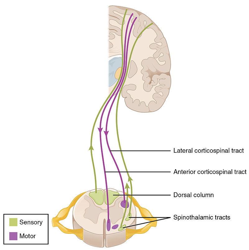
Diagram of the columns and of the course of the fibers in the spinal cord. Sensory synapses occur in the dorsal spinal cord (above in this image), and motor nerves leave through the ventral (as well as lateral) horns of the spinal cord as seen below in the image.
Schematic image showing the locations of a few tracts of the spinal cord.
Illustration from Anatomy & Physiology, Connexions Web site. http://cnx.org/content/col11496/1.6/, Jun 19, 2013..
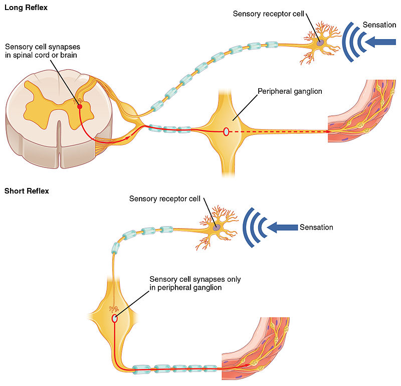
Reflexes may also occur without engaging more than one neuron of the CNS as in the below example of a short reflex.
Illustration from Anatomy & Physiology, Connexions Web site. http://cnx.org/content/col11496/1.6/, Jun 19, 2013.
Different ways in which the CNS can be activated without engaging the cortex, and making us aware of the actions. The above example shows the process in which the pupil dilates during dim light, activating neurons in the spinal cord. The second example shows the constriction of the pupil as a result of the activation of the Eddinger-Westphal nucleus (a cerebral ganglion)..
Illustration from Anatomy & Physiology, Connexions Web site. http://cnx.org/content/col11496/1.6/, Jun 19, 2013.
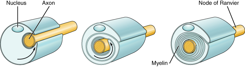
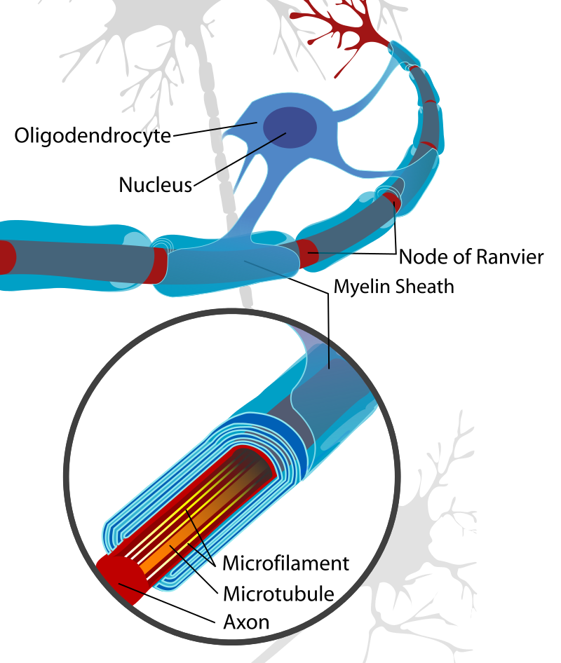
A peripheral nerve myelinated by Schwann cells (left) and a CNS neuron myelinated by an oligodendrocyte (right).
A peripheral nerve myelinated by Schwann cells (left) and a CNS neuron myelinated by an oligodendrocyte (right).
A neuron cell diagram, cropped to show oligodendrocyte and myelin sheath.
A map over the different structures of the nervous systems in the body, showing the CNS, PNS,autonomic nervous system, and enteric nervous system.
Version 8.25 from the Textbook OpenStax Anatomy and Physiology Published May 18, 2016.
CNS as seen in a median section of a 5 week old embryo.
An anatomical illustration from Sobotta's Human Anatomy 1908.
CNS seen in a median section of a 3 month old embryo.
An anatomical illustration from Sobotta's Human Anatomy 1908.
Diagram depicting the main subdivisions of the embryonic vertebrate brain, later forming forebrain,midbrain and hindbrain.
Diagram depicting the main subdivisions of the embryonic vertebrate brain. The neural tube differentiates into forebrain, midbrain and hindbrain structures.
Development of the neural tube.
Development of the neural tube in human embryos (Prentiss-Arey). A. An early embryo (Keibel) B. at 2 mm. (Graf Spee) C. at 2 mm. (Mall) D. at 2.7 mm (Kollmann). This is a scan of Figure 6 of the book "The anatomy of the nervous system" by Stephen Walter Ranson, with the labels redrawn..
The lancelet, regarded an archetypal vertebrate, lacking a true brain.
A Lancelet (or Amphioxus) specimen —Subphylum: Cephalochordata— collected in coarse sand sediments (600 µm) on the Belgian continental shelf. Total Length: approximately 22 mm. Geo-location not applicable as the picture was taken in the laboratory.
Spindle diagram of the evolution of vertebrates.
Evolution of Vertebrate from the Cambrium to the present at a class level as a traditional spindle diagram. The width of the spindle represents the number of families as a rough estimate of diversity. The diagram is based on Benton, M. J. (1998) The quality of the fossil record of vertebrates. P. 269–303, in Donovan, S. K. and Paul, C. R. C. (eds), The adequacy of the fossil record, Fig. 2. Wiley, New York, 312 p. The figures representing classes are (from left): Agnatha, Chondrichthyes, Osteichthyes, Amphibia, Reptilia, Aves and Mammalia. The two extinct classes are Placodermi and Acanthodii. All classes interpreted traditionally. Bentons notes to his own tree: Number of families is an imperfect measure of diversity. Reptilia in particular should probably have been shown as far more diverse in the Mesozoic..
Chlorophyll (also chlorophyl) is any of several related green pigments found in the mesosomes of cyanobacteria and in the chloroplasts of algae and plants. Its name is derived from the Greek words χλωρός, khloros ("pale green") and φύλλον, phyllon ("leaf"). Chlorophyll is essential in photosynthesis, allowing plants to absorb energy from light.
Chlorophylls absorb light most strongly in the blue portion of the electromagnetic spectrum as well as the red portion. Conversely, it is a poor absorber of green and near-green portions of the spectrum, which it reflects, producing the green color of chlorophyll-containing tissues. Two types of chlorophyll exist in the photosystems of green plants: chlorophyll a and b. (W)
Seen through a microscope, chlorophyll is concentrated within organisms in structures called chloroplasts – shown here grouped inside plant cells.
Chloroplasts visible in the cells of Bryum capillare, a type of moss.
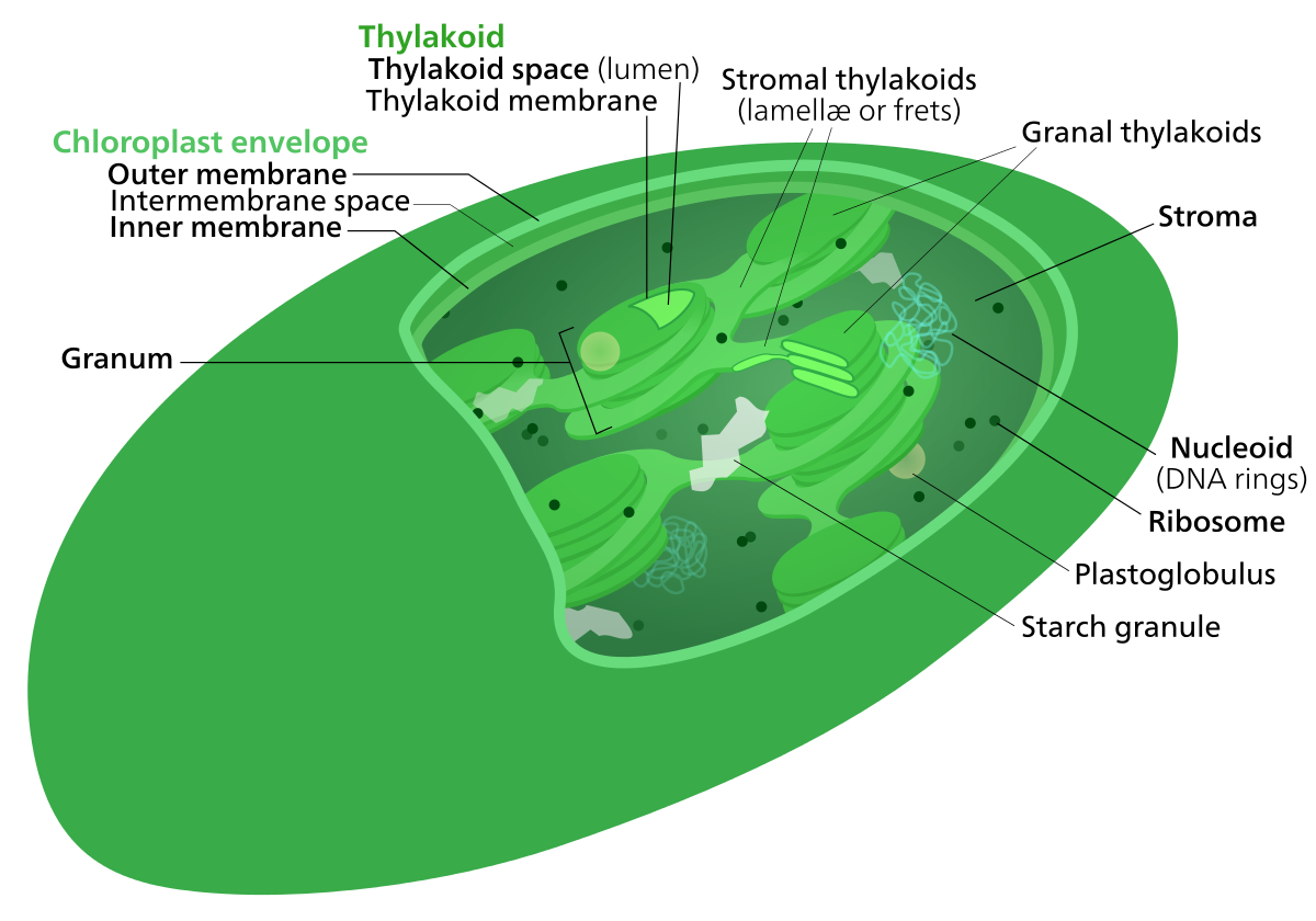
Chloroplast ultrastructure(interactive diagram) Chloroplasts have at least three distinct membrane systems, and a variety of things can be found in their stroma.
chromatin
Chromatin is a complex of DNA and proteinfound in eukaryotic cells. Its primary function is packaging long DNA molecules into more compact, denser structures. This prevents the strands from becoming tangled and also plays important roles in reinforcing the DNA during cell division,preventing DNA damage, and regulating gene expression and DNA replication. During mitosis and meiosis, chromatin facilitates proper segregation of the chromosomes in anaphase; the characteristic shapes of chromosomes visible during this stage are the result of DNA being coiled into highly condensed chromatin. (W)
A cartoon representation of the nucleosome structure. From PDB:1KX5.
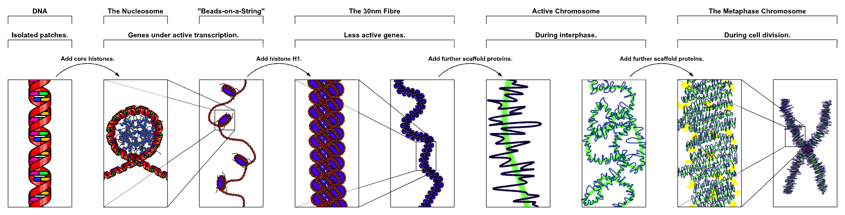
Basic units of chromatin structure.
The major structures in DNA compaction DNA, the nucleosome, the 10 nm “beads-on-a-string” fibre, the 30 nm chromatin fibre and the metaphase chromosome
🔎
Cnidaria is a phylum under kingdom Animalia containing over 11,000 species of aquatic animals found both in freshwater and marine environments, predominantly the latter.
Their distinguishing feature is cnidocytes, specialized cells that they use mainly for capturing prey. Their bodies consist of mesoglea, a non-living jelly-like substance, sandwiched between two layers of epithelium that are mostly one cell thick. (W)
Collective cell migration describes the movements of group of cells and the emergence of collective behavior from cell-environment interactions and cell-cell communication. Collective cell migration is an essential process in the lives of multicellular organisms, e.g. embryonic development,wound healing and cancer spreading (metastasis). Cells can migrate as a cohesive group (e.g. epithelial cells) or have transient cell-cell adhesion sites (e.g. mesenchymal cells). They can also migrate in different modes like sheets, strands, tubes, and clusters. While single-cell migration has been extensively studied, collective cell migration is a relatively new field with applications in preventing birth defects or dysfunction of embryos. It may improve cancer treatment by enabling doctors to prevent tumors from spreading and forming new tumors.(W)
CRISPR
CRISPR (clustered regularly interspaced short palindromic repeats) is a family of DNA sequences found in the genomes of prokaryotic organisms such as bacteria and archaea. These sequences are derived from DNA fragments of bacteriophages that had previously infected the prokaryote. They are used to detect and destroy DNA from similar bacteriophages during subsequent infections. Hence these sequences play a key role in the antiviral (i.e. anti-phage) defense system of prokaryotes.
The CRISPR-Cas system is a prokaryotic immune system that confers resistance to foreign genetic elements such as those present within plasmids and phages and provides a form of acquired immunity. RNA harboring the spacer sequence helps Cas (CRISPR-associated) proteins recognize and cut foreign pathogenic DNA. Other RNA-guided Cas proteins cut foreign RNA. CRISPR are found in approximately 50% of sequenced bacterial genomes and nearly 90% of sequenced archaea.
These systems have created CRISPR gene editing that commonly utilizes the cas9 gene. This editing process has a wide variety of applications including basic biological research, development of biotechnology products, and treatment of diseases. (W)
Diagram of the CRISPR prokaryotic antiviral defense mechanism.
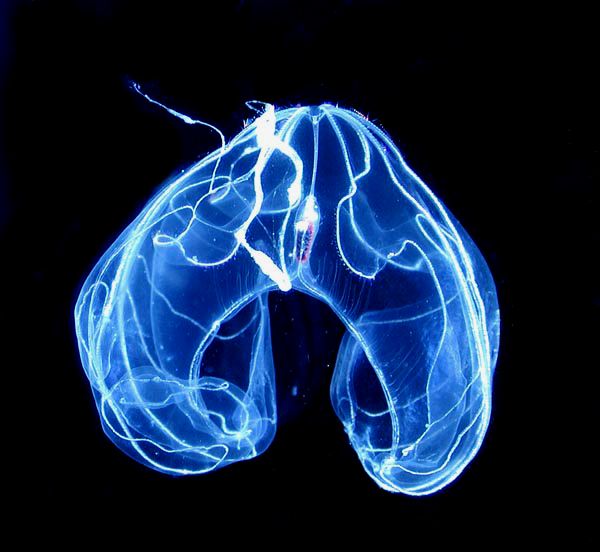
ctenophora
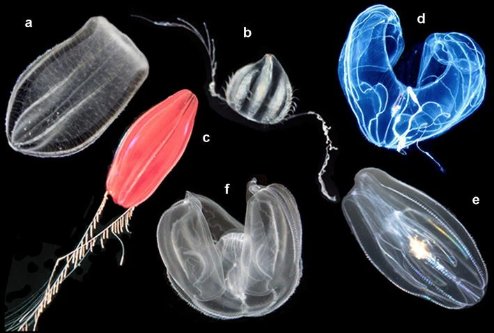
Ctenophora (singular ctenophore, from Ancient Greek:κτείς, romanized:kteis, lit. 'comb' and φέρω, pherō, 'to carry'; commonly known as comb jellies) comprise a phylum of invertebrate animals that live in marine waters worldwide. They are notable for the groups of cilia they use for swimming (commonly referred to as "combs"), and they are the largest animals to swim with the help of cilia. Depending on the species, adult ctenophores range from a few millimeters to 1.5 m (4 ft 11 in) in size. Only 100 to 150 species have been validated, and possibly another 25 have not been fully described and named. The textbook examples are cydippids with egg-shaped bodies and a pair of retractable tentacles fringed with tentilla ("little tentacles") that are covered with colloblasts, sticky cells that capture prey. Their bodies consist of a mass of jelly, with a layer two cells thick on the outside, and another lining the internal cavity. The phylum has a wide range of body forms, including the egg-shaped cydippids with retractable tentacles that capture prey, the flat generally combless platyctenids, and the large-mouthed beroids, which prey on other ctenophores. (W)
Enlarge image for index to numbers. Haeckelia rubra (Victor Carus, 1862) = Haeckelia rubra (Gegenbaur, Kölliker & Müller, 1853), from above Haeckelia rubra (Victor Carus, 1862) = Haeckelia rubra (Gegenbaur, Kölliker & Müller, 1853), from the side Hormiphora foliosa (Haeckel) = Hormiphora sp., from the side Callianira bialata (Delle Chiaje) = Callianira bialata Delle Chiaje, 1841, from the side Tinerfe cyanea (Chun) = Tinerfe cyanea (Chun, 1889), from the side Lampetia pancerina (Chun) = Lampea pancerina (Chun, 1879), from the side.
Bathocyroe fosteri a common but fragile deep-sea lobate, oriented mouth down.
Undescribed deep-sea species known as "Tortugas red", with trailing tentacles and clearly visible sidebranches, or tentilla.
cyanobacteria
Cyanobacteria, also known as Cyanophyta, are a phylum consisting of both free-living photosynthetic bacteria and the endosymbioticplastids that are present in the Archaeplastida autotrophic eukaryotes. The plastids are a sister group to the free-living Gloeomargarita. Cyanobacteria commonly obtain their energy through oxygenic photosynthesis. The oxygen gas in the atmosphere of earth is produced by cyanobacteria of this phylum, either as free-living bacteria or as the endosymbiotic plastids. The name cyanobacteria comes from the color of the bacteria (Greek:κυανός, romanized:kyanós, lit. 'blue'). Cyanobacteria, which are prokaryotes, are also called "blue-green algae", though some modern botanists restrict the term algae to eukaryotes. Cyanobacteria appear to have originated in freshwater or a terrestrial environment. (W)
Light microscope view of cyanobacteria from a microbial mat.
The cytosol, also known as intracellular fluid (ICF) or cytoplasmic matrix, or groundplasm, is the liquid found inside cells.It is separated into compartments by membranes. For example, the mitochondrial matrix separates the mitochondrion into many compartments.
In the eukaryotic cell, the cytoplasm is surrounded by the cell membrane and is part of the cytoplasm, which also comprises the mitochondria, plastids, and other organelles (but not their internal fluids and structures); the cell nucleus is separate. The cytosol is thus a liquid matrix around the organelles. In prokaryotes, most of the chemical reactions of metabolism take place in the cytosol, while a few take place in membranes or in the periplasmic space. In eukaryotes, while many metabolic pathways still occur in the cytosol, others take place within organelles.
The cytosol is a complex mixture of substances dissolved in water. Although water forms the large majority of the cytosol, its structure and properties within cells is not well understood. The concentrations of ions such as sodium and potassium are different in the cytosol than in the extracellular fluid; these differences in ion levels are important in processes such as osmoregulation,cell signaling, and the generation of action potentials in excitable cells such as endocrine, nerve and muscle cells. The cytosol also contains large amounts of macromolecules, which can alter how molecules behave, through macromolecular crowding.
A cytotoxic T cell (also known as TC, cytotoxic T lymphocyte, CTL, T-killer cell, cytolytic T cell, CD8+ T-cell or killer T cell) is a T lymphocyte (a type of white blood cell) that kills cancer cells, cells that are infected (particularly with viruses), or cells that are damaged in other ways.
Most cytotoxic T cells express T-cell receptors (TCRs) that can recognize a specific antigen. An antigen is a molecule capable of stimulating an immune response and is often produced by cancer cells or viruses. Antigens inside a cell are bound to class I MHC molecules, and brought to the surface of the cell by the class I MHC molecule, where they can be recognized by the T cell. If the TCR is specific for that antigen, it binds to the complex of the class I MHC molecule and the antigen, and the T cell destroys the cell.
In order for the TCR to bind to the class I MHC molecule, the former must be accompanied by a glycoprotein called CD8, which binds to the constant portion of the class I MHC molecule. Therefore, these T cells are called CD8+ T cells. (W)
Antigen presentation stimulates T cells to become either "cytotoxic" CD8+ cells or "helper" CD4+ cells.
Antigen presentation stimulates T cells to activate "cytotoxic" CD8+ cells or "helper" CD4+ cells. Cytotoxic cells directly attack other cells carrying certain foreign or abnormal molecules on their surfaces. Helper T cells, or Th cells, coordinate immune responses by communicating with other cells. In most cases, T cells only recognize an antigen if it is carried on the surface of a cell by one of the body’s own MHC, or major histocompatibility complex, molecules.
Development of single positive T cells in the thymus.
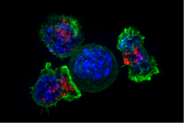
In this immunofluorescence image, a group of killer T cells (outer three) is engaging a cancer cell (centered one). A patch of signaling molecules (pink) that gathers at the site of cell-cell contact indicates that the CTL has identified a target. Lytic granules (red) that contain cytotoxic components then travel along the microtubule cytoskeleton (green) to the contact site and are secreted, thus killing the target.
Superresolution image of a group of killer T cells (green and red) surrounding a cancer cell (blue, center). When a killer T cell makes contact with a target cell, the killer cell attaches and spreads over the dangerous target. The killer cell then uses special chemicals housed in vesicles (red) to deliver the killing blow. This event has thus been nicknamed “the kiss of death”. After the target cell is killed, the killer T cells move on to find the next victim.
d
Darwinism
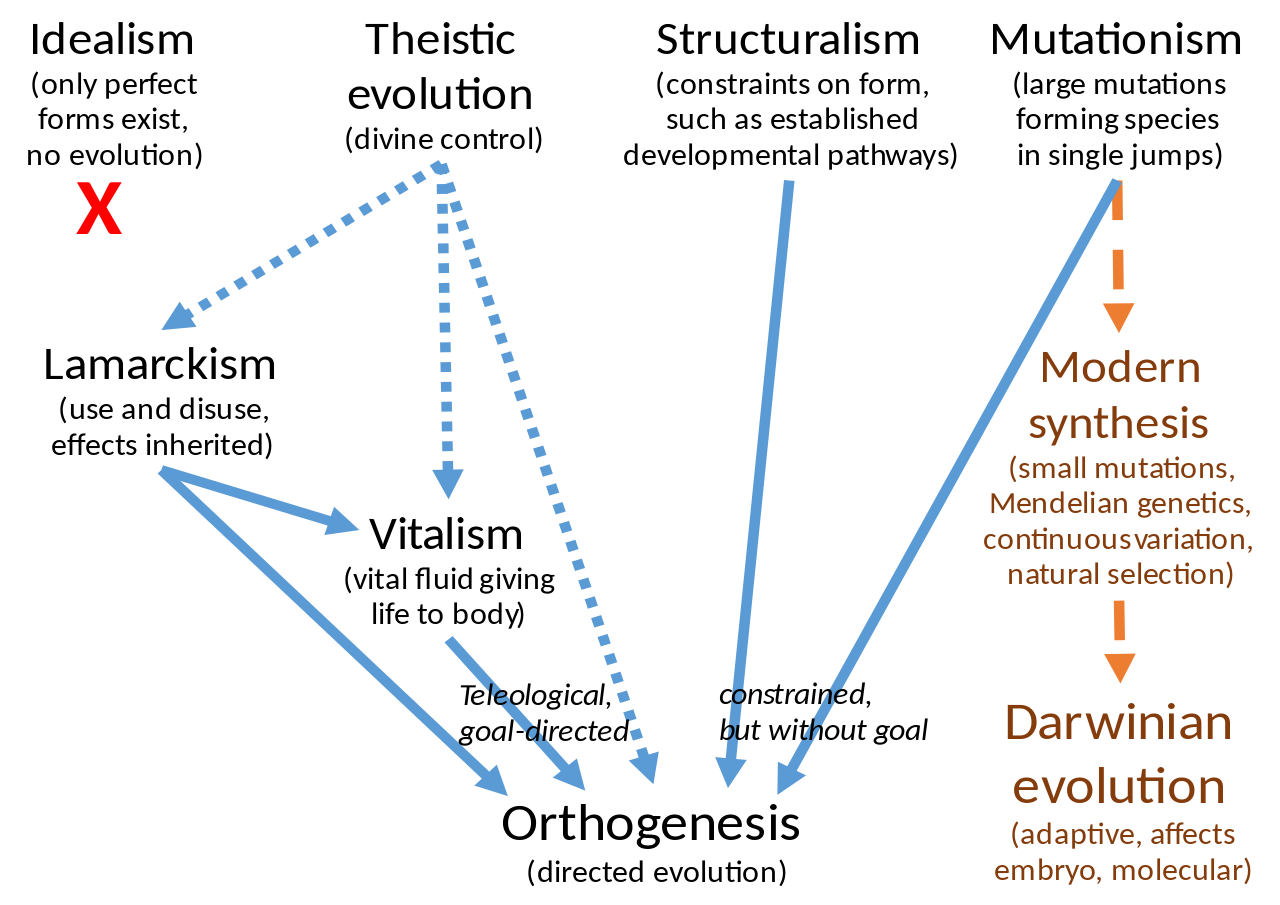
Darwinism is a theory of biologicalevolution developed by the English naturalist Charles Darwin (1809–1882) and others, stating that all species of organisms arise and develop through the natural selection of small, inherited variations that increase the individual's ability to compete, survive, and reproduce. Also called Darwinian theory, it originally included the broad concepts of transmutation of species or of evolution which gained general scientific acceptance after Darwin published On the Origin of Species in 1859, including concepts which predated Darwin's theories. English biologist Thomas Henry Huxley coined the term Darwinism in April 1860. (W)
Darwinism, the eclipse of
Julian Huxley used the phrase “the eclipse of Darwinism” to describe the state of affairs prior to what he called the modern synthesis, when evolution was widely accepted in scientific circles but relatively few biologists believed that natural selection was its primary mechanism. Historians of science such as Peter J. Bowler have used the same phrase as a label for the period within the history of evolutionary thought from the 1880s to around 1920, when alternatives to natural selection were developed and explored—as many biologists considered natural selection to have been a wrong guess on Charles Darwin's part, or at least as of relatively minor importance.An alternative term, the interphase of Darwinism, has been proposed to avoid the largely incorrect implication that the putative eclipse was preceded by a period of vigorous Darwinian research.
Theistic evolution was the belief that God directly guided evolution.
Neo-Lamarckism was the idea that evolution was driven by the inheritance of characteristics acquired during the life of the organism.
Orthogenesis was the belief that organisms were affected by internal forces or laws of development that drove evolution in particular directions
Mutationism was the idea that evolution was largely the product of mutations that created new forms or species in a single step.
Theistic evolution largely disappeared from the scientific literature by the end of the 19th century as direct appeals to supernatural causes came to be seen as unscientific. The other alternatives had significant followings well into the 20th century; mainstream biology largely abandoned them only when developments in genetics made them seem increasingly untenable, and when the development of population genetics and the modern synthesis demonstrated the explanatory power of natural selection.Ernst Mayr wrote that as late as 1930 most textbooks still emphasized such non-Darwinian mechanisms. (W)
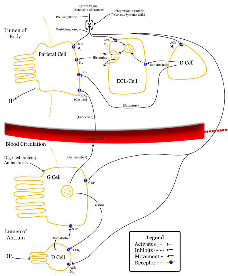
Delta cells (δ-cells or D cells) are somatostatin-producing cells. They can be found in the stomach,intestine and the pancreatic islets. In rodents, delta-cells are located in the periphery of the islets; in humans the islet architecture is generally less organized and delta-cells are frequently observed inside the islets as well. In both species, the peptide hormone Urocortin3 (Ucn3) is a major local signal that is released from beta cells (and alpha cells in primates) to induce the local secretion of somatostatin. It has also been suggested that somatostatin may be implicated in insulin-induced hypoglycaemia through a mechanism involving SGLT-2 receptors. Ghrelin can also strongly stimulate somatostatin secretion, thus indirectly inhibiting insulin release. Viewed under an electron microscope, delta-cells can be identified as cells with smaller and slightly more compact granules than beta cells. (W)
Diagram summarising control of stomach acid secretion. There is an blank version of this (ie. no text) ready for other languages here. If you want the Photoshop .pdf file directly just let me know on my talk page (It's quite large so wouldn't want to host on wikipedia).
Dendritic cells are present in those tissues that are in contact with the external environment, such as the skin (where there is a specialized dendritic cell type called the Langerhans cell) and the inner lining of the nose,lungs,stomach and intestines. They can also be found in an immature state in the blood. Once activated, they migrate to the lymph nodes where they interact with T cells and B cells to initiate and shape the adaptive immune response. At certain development stages they grow branched projections, the dendrites that give the cell its name (δένδρον or déndron being Greek for 'tree'). While similar in appearance, these are structures distinct from the dendrites of neurons. Immature dendritic cells are also called veiled cells, as they possess large cytoplasmic 'veils' rather than dendrites. (W)
Dendritic cells in skin.
Section of skin showing large numbers of dendritic (Langerhans) cells in the epidermis. M. ulcerans infection, S100 immunoperoxidase stain.
Title: Dendritic cell revealed Description: Artistic rendering of the surface of a human dendritic cell illustrating sheet-like processes that fold back onto the membrane surface. When exposed to HIV, these sheets entrap viruses in the vicinity and focus them to contact zones with T-cells targeted for infection. These studies were carried out using ion abrasion scanning electron microscopy, a new technology we have been developing and applying for 3D cellular imaging. Categories: Research in NIH Labs and Clinics Type: Color, Diagram Source: National Cancer Institute (NCI) Creator: Don Bliss, Sriram Subramaniam Date Created: Unknown Date Added: 5/24/2012 Reuse Restrictions: None - This image is in the public domain and can be freely reused.
A dendritic cell.
A screen clip from a video included in the journal article “Environmental Dimensionality Controls the Interaction of Phagocytes with the Pathogenic Fungi Aspergillus fumigatus and Candida albicans” A well resolved dendritic cell drags a conidium through a distance of up to 9 μm. The conidium, however, is not phagocytosed by the cell.
diatom
Diatoms (diá-tom-os 'cut in half', from diá, 'through' or 'apart'; and the root of tém-n-ō, 'I cut'.) are a major group of algae, specifically microalgae, found in the oceans, waterways and soils of the world. Living diatoms make up a significant portion of the Earth's biomass: they generate about 20 to 50 percent of the oxygen produced on the planet each year, take in over 6.7 billion metric tons of silicon each year from the waters in which they live, and constitute nearly half of the organic material found in the oceans. The shells of dead diatoms can reach as much as a half-mile (800 m) deep on the ocean floor, and the entire Amazon basin is fertilized annually by 27 million tons of diatom shell dust transported by transatlantic winds from the African Sahara, much of it from the Bodélé Depression, which was once made up of a system of fresh-water lakes. (W)
Assorted diatoms as seen through a microscope. These specimens were living between crystals of annual sea ice in McMurdo Sound, Antarctica. Image digitized from original 35mm Ektachrome slide. These tiny phytoplankton are encased within a silicatecell wall.
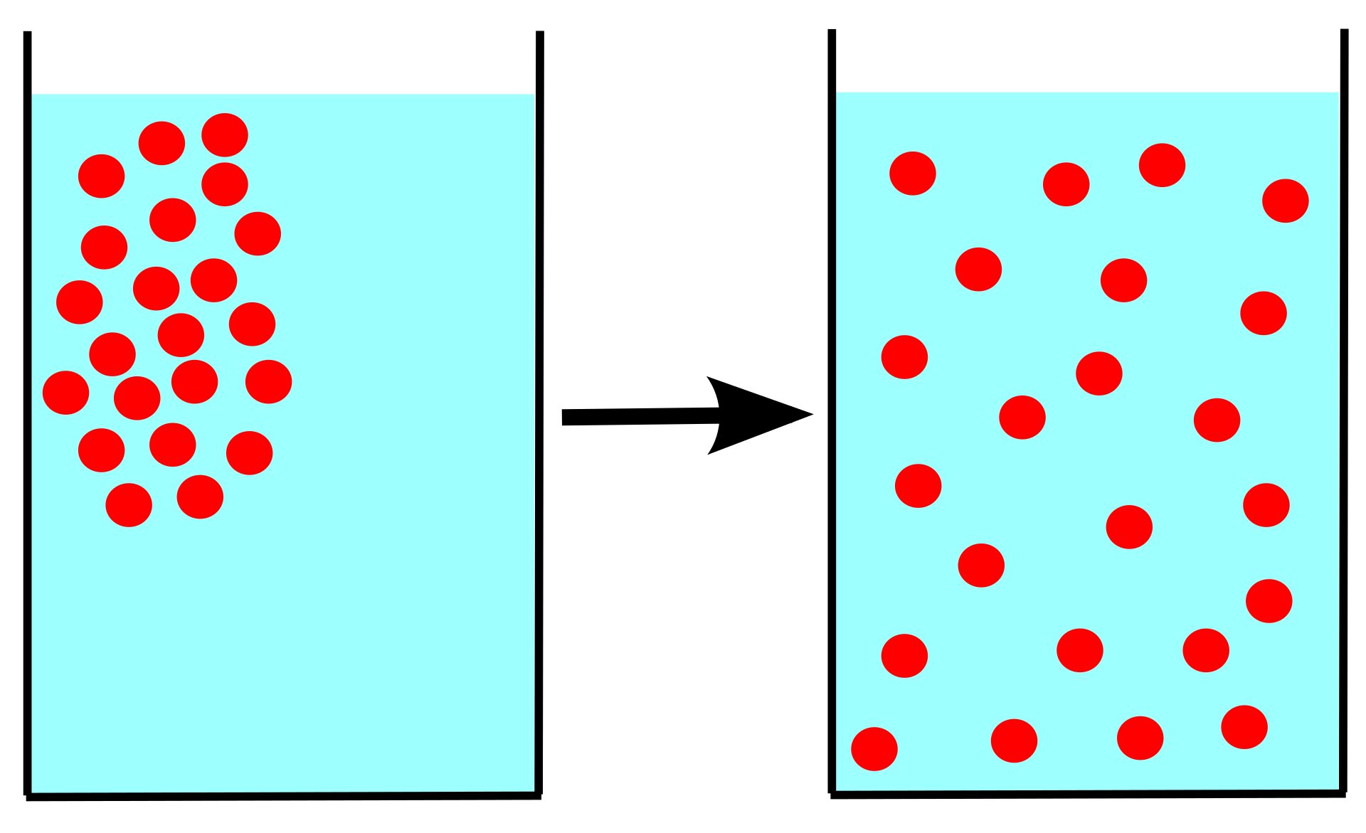
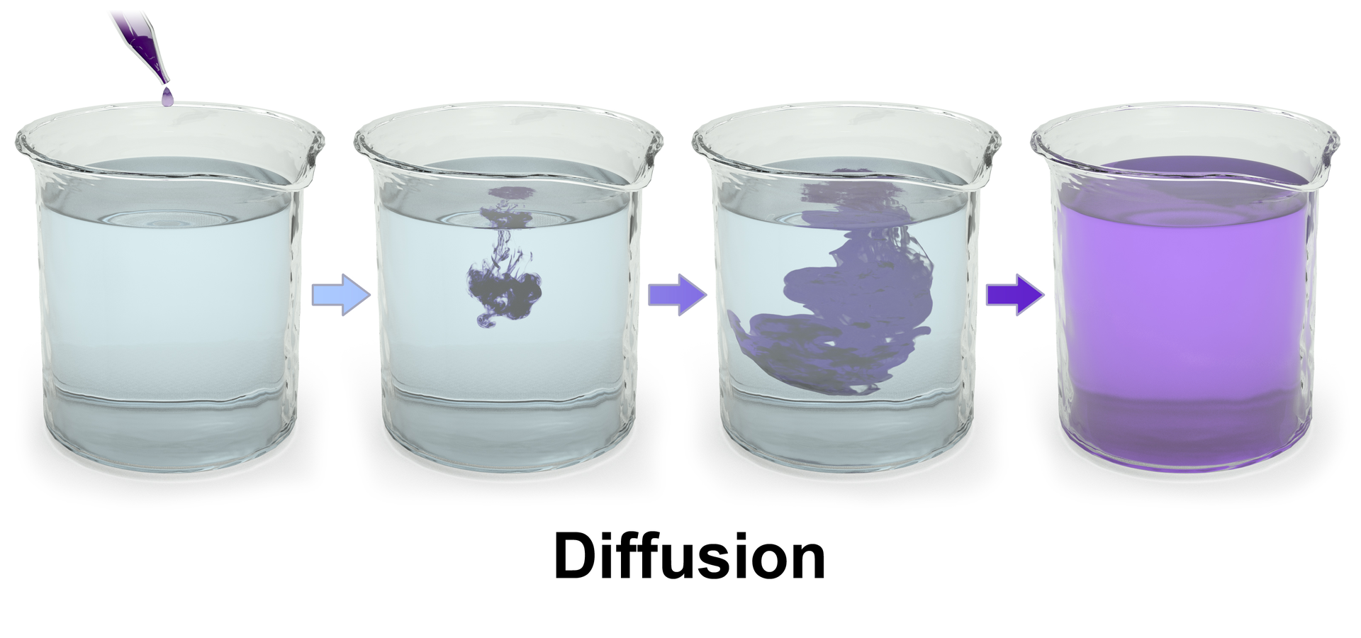
diffusion
Diffusion is the net movement of anything (for example, atom, ions, molecules) from a region of higher concentration to a region of lower concentration. Diffusion is driven by a gradient in concentration.
The concept of diffusion is widely used in many fields, including physics (particle diffusion),chemistry,biology,sociology,economics, and finance (diffusion of people, ideas, and price values). The central idea of diffusion, however, is common to all of these: an object (for example, atom, idea, etc.) undergoing diffusion spreads out from a point or location at which there is a higher concentration of that object.
The word diffusion derives from the Latin word, diffundere, which means "to spread out."(W)
Some particles are dissolved in a glass of water. At first, the particles are all near one top corner of the glass. If the particles randomly move around ("diffuse") in the water, they eventually become distributed randomly and uniformly from an area of high concentration to an area of low concentration, and organized (diffusion continues, but with no net flux).
Three-dimensional rendering of diffusion of purple dye in water.
Diploid cells have two homologous copies of each chromosome, usually one from the mother and one from the father. All or nearly all mammals are diploid organisms. The suspected tetraploid (possessing four chromosome sets) plains viscacha rat (Tympanoctomys barrerae) and golden viscacha rat (Pipanacoctomys aureus) have been regarded as the only known exceptions (as of 2004). However, some genetic studies have rejected any polyploidism in mammals as unlikely, and suggest that amplification and dispersion of repetitive sequences best explain the large genome size of these two rodents. All normal diploid individuals have some small fraction of cells that display polyploidy.Human diploid cells have 46 chromosomes (the somatic number, 2n) and human haploid gametes (egg and sperm) have 23 chromosomes (n). Retroviruses that contain two copies of their RNA genome in each viral particle are also said to be diploid. Examples include human foamy virus,human T-lymphotropic virus, and HIV(W)
A haploid set that consists of a single complete set of chromosomes (equal to the monoploid set), as shown in the picture above, must belong to a diploid species. If a haploid set consists of two sets, it must be of a tetraploid (four sets) species.


.jpg)

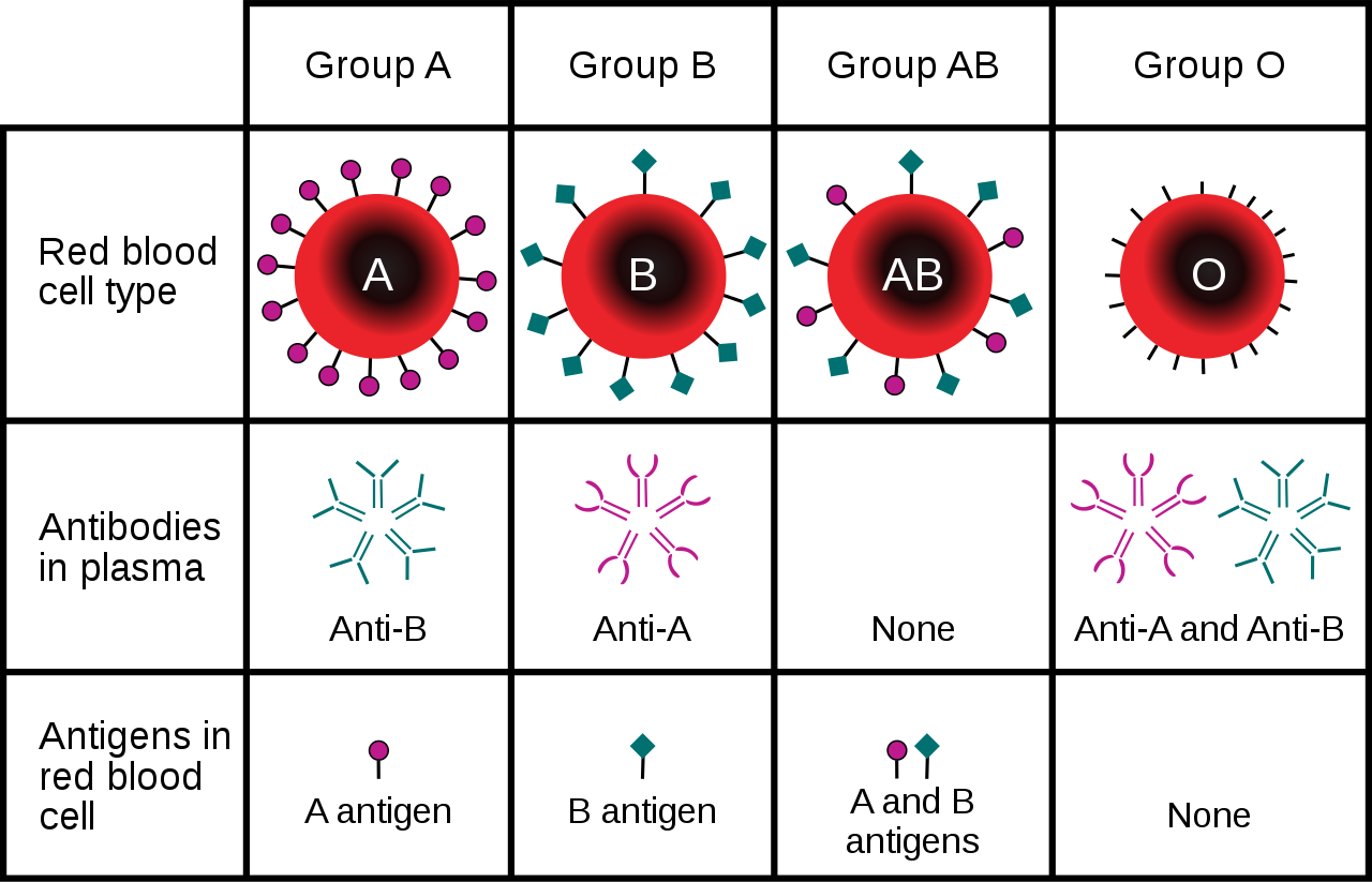


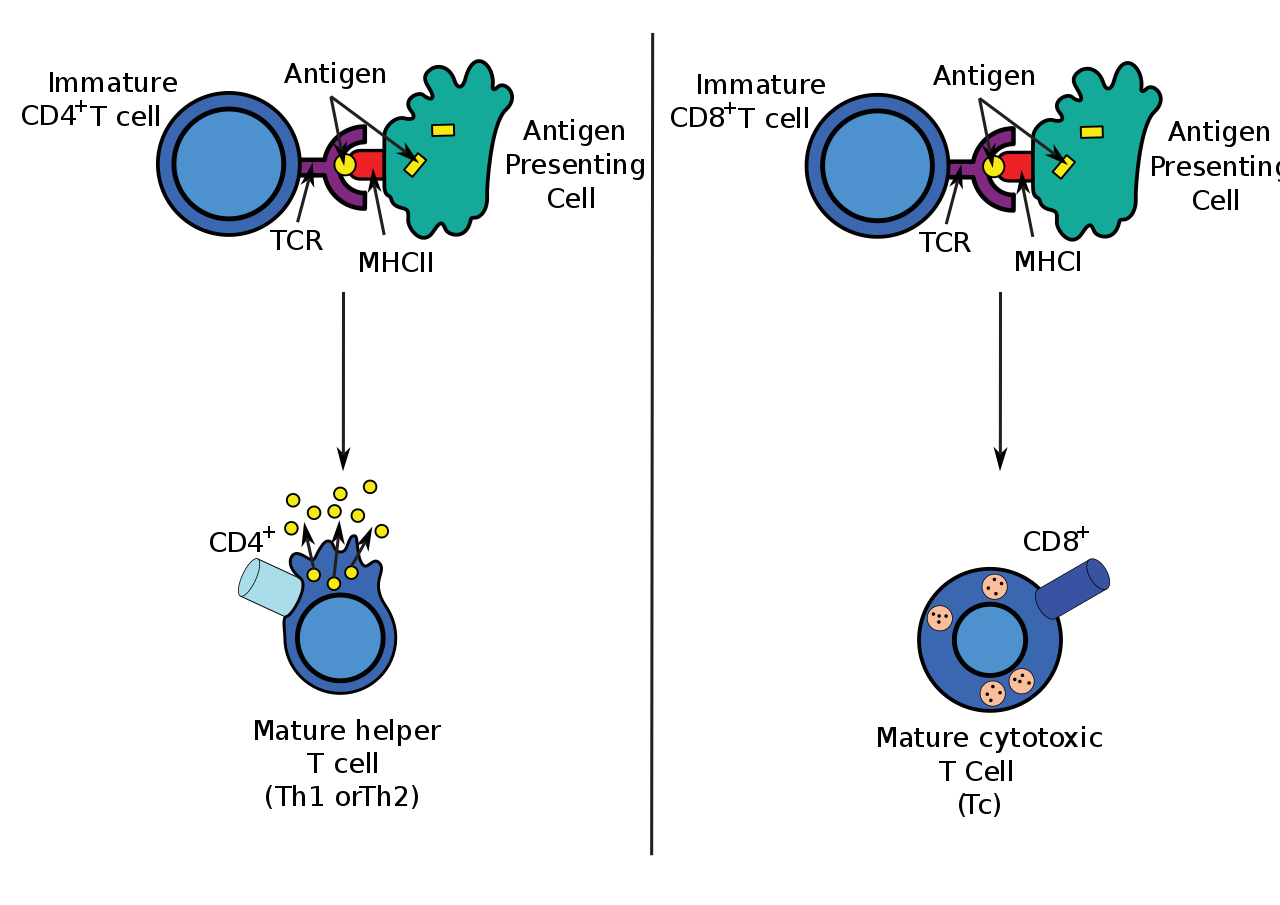


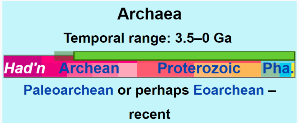


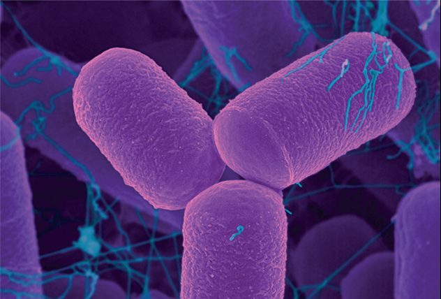
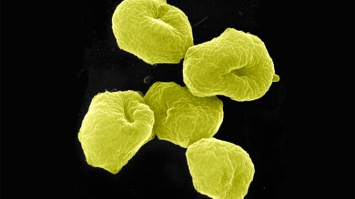
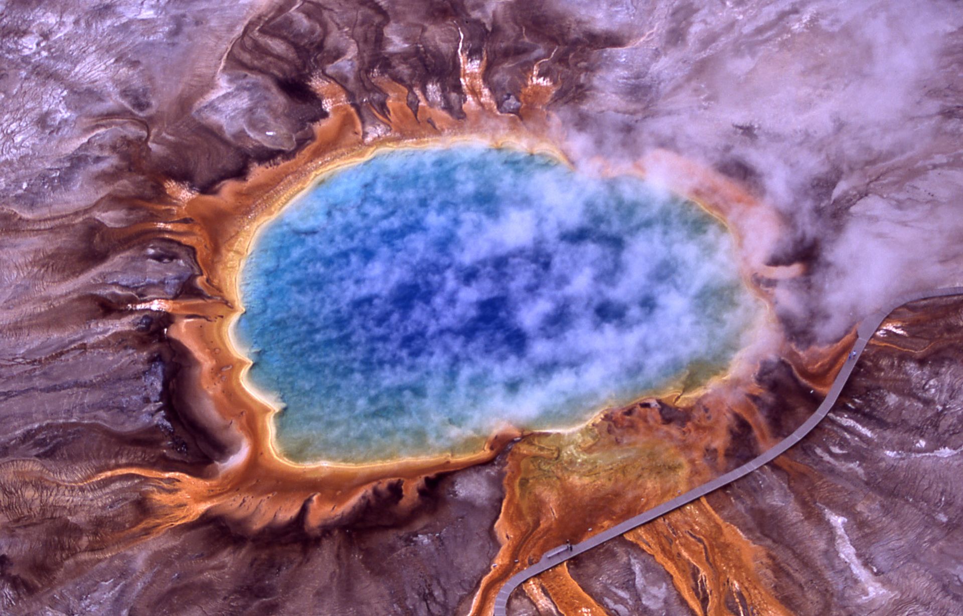

.png)

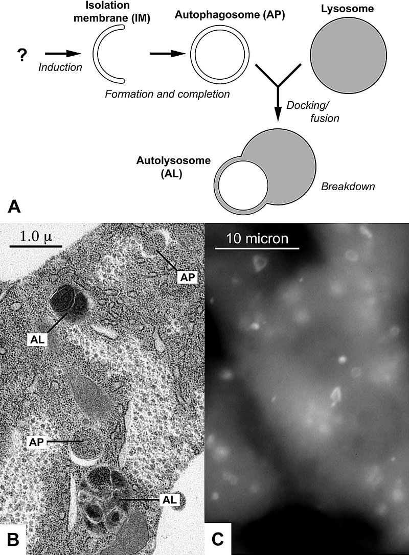
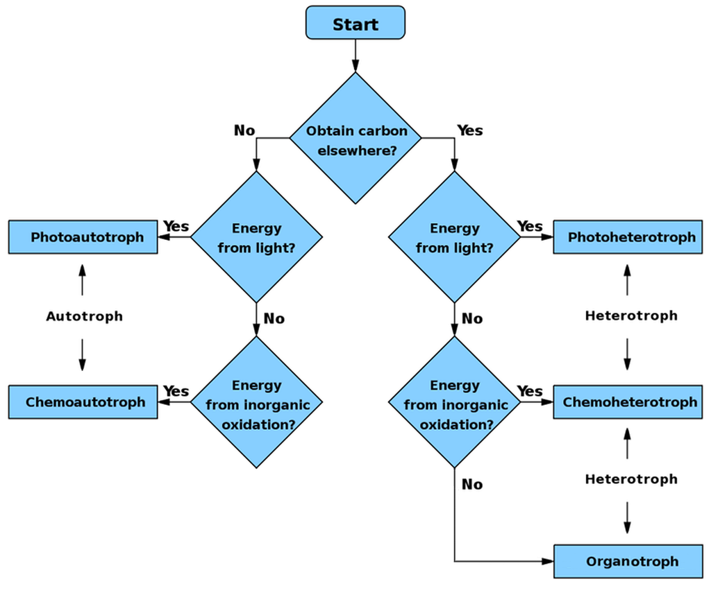


.jpg)


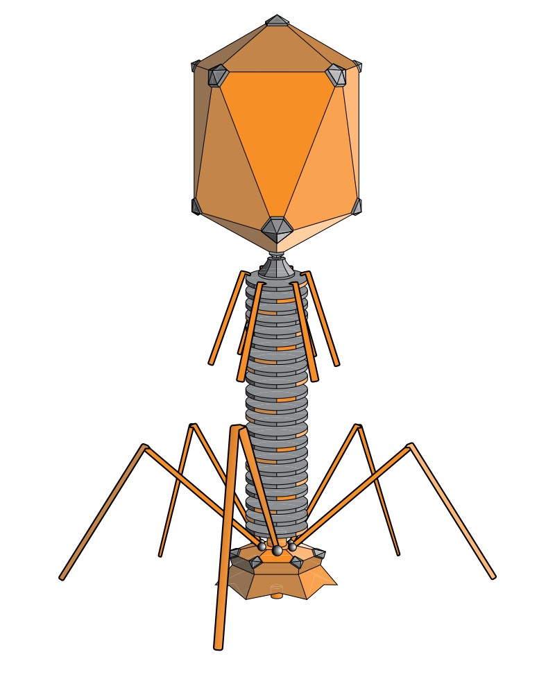

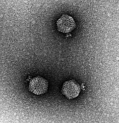


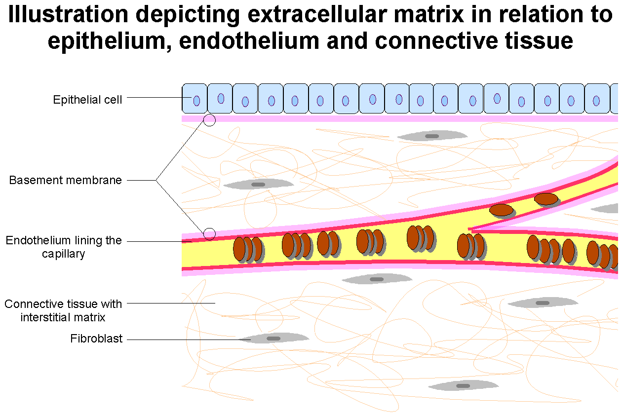


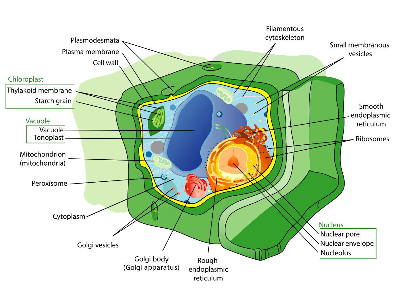
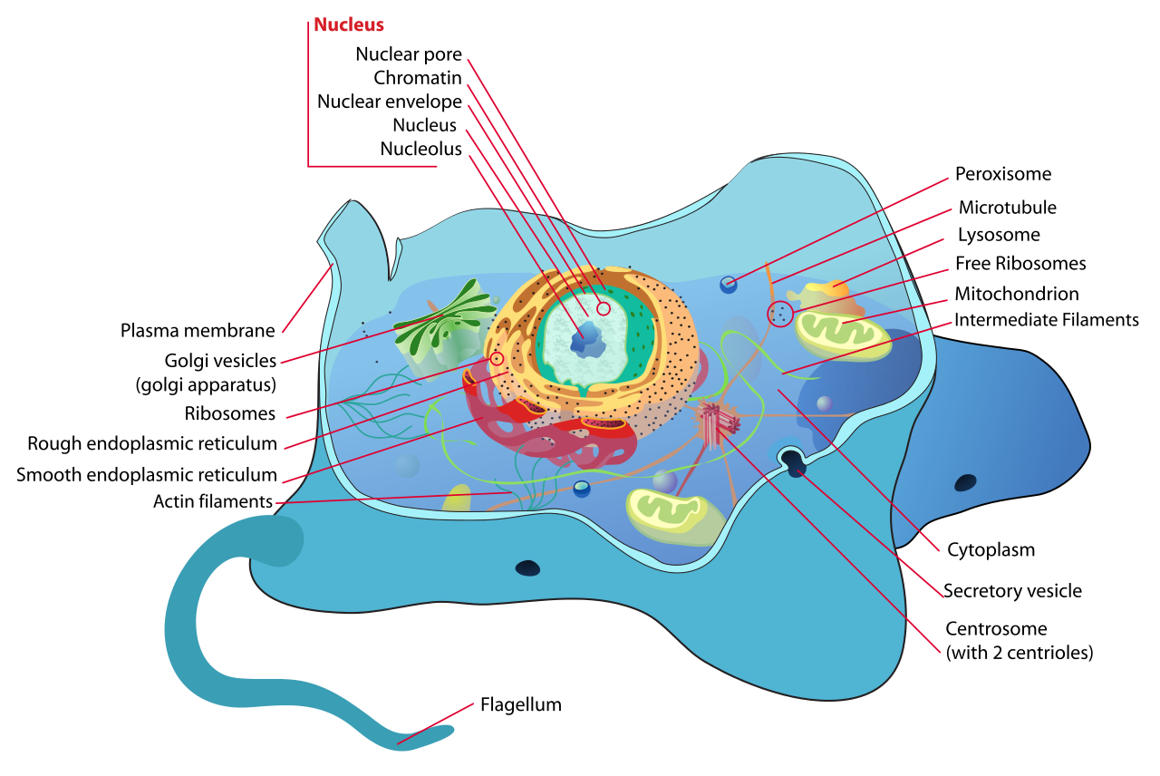
.png)
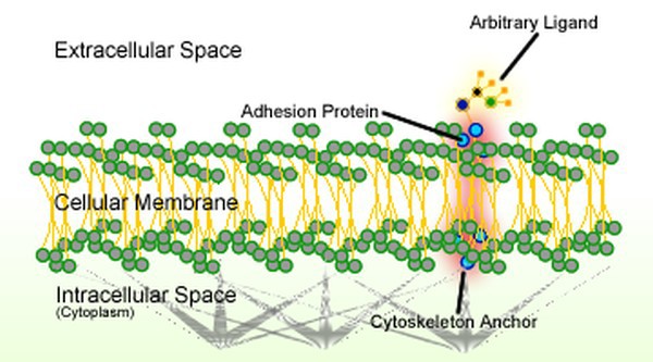


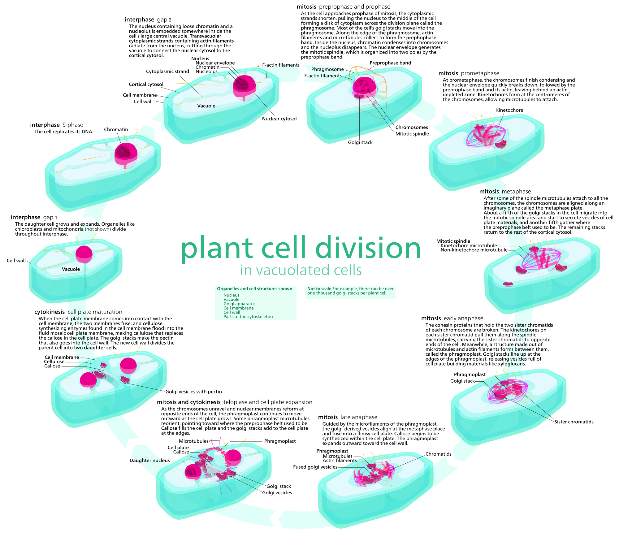


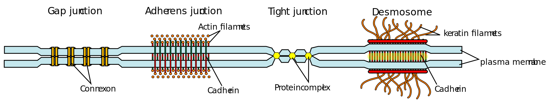


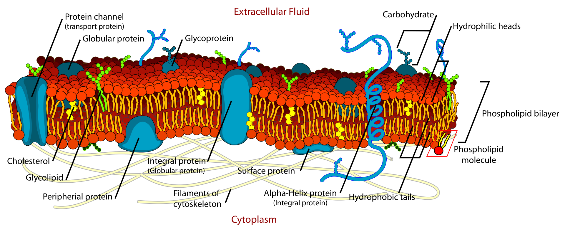

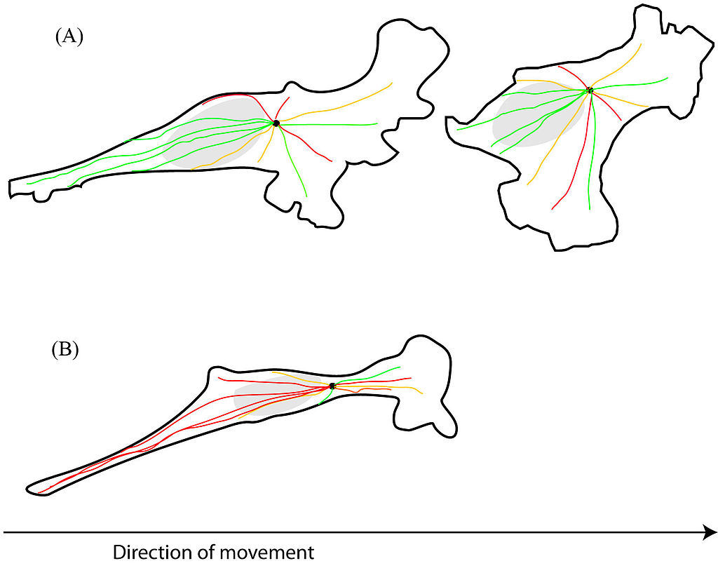

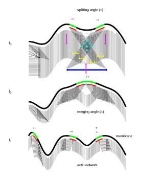


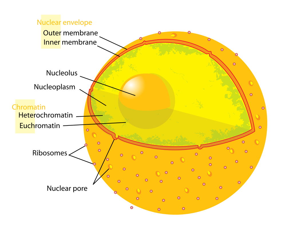

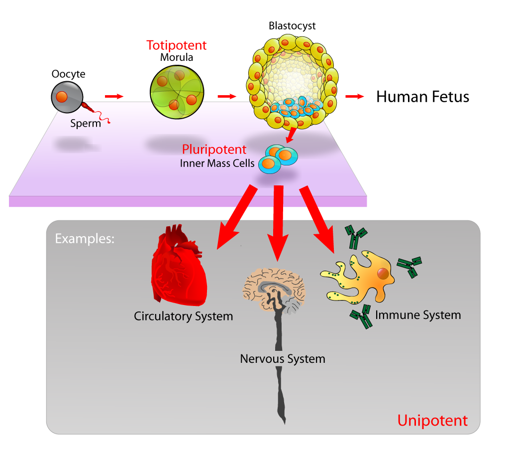

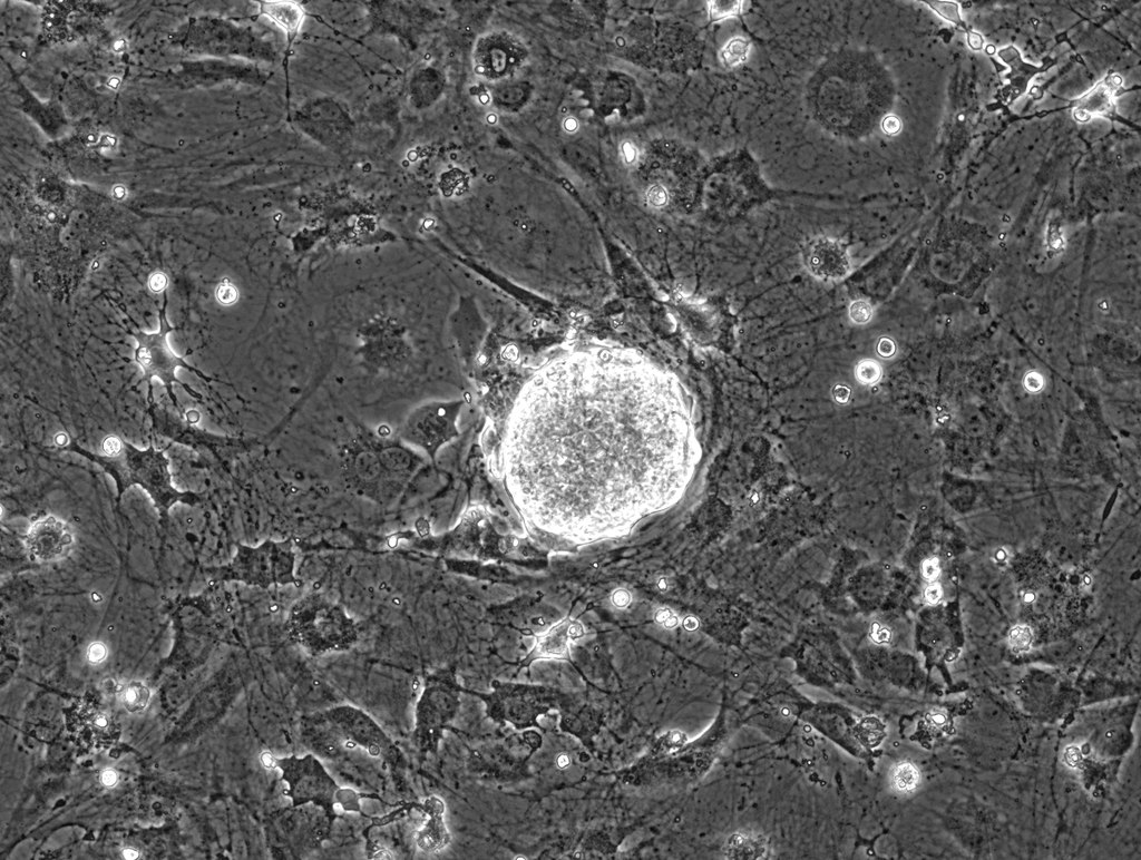
_diagram_en.png)